
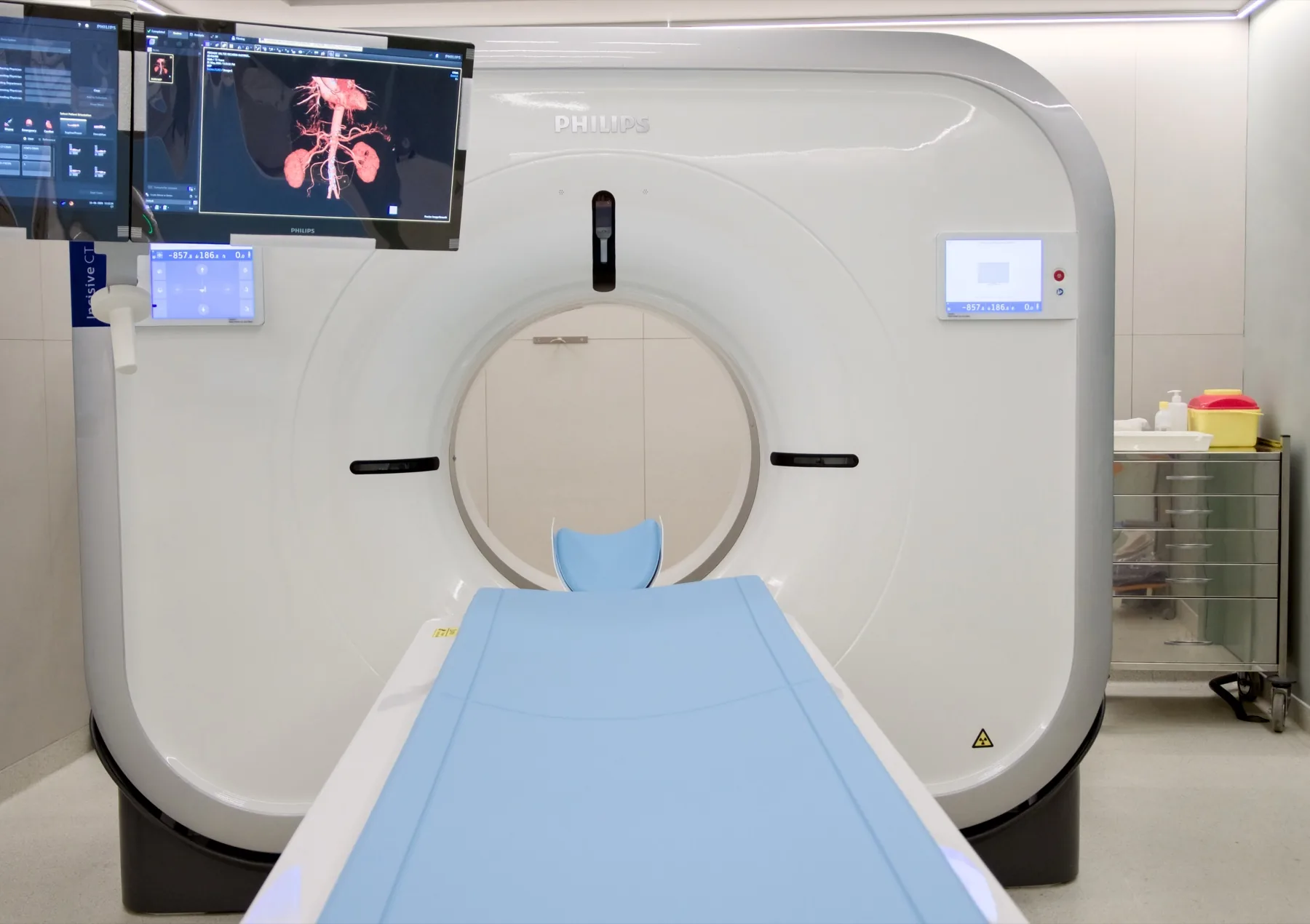
What is assessed in a CT enterography?
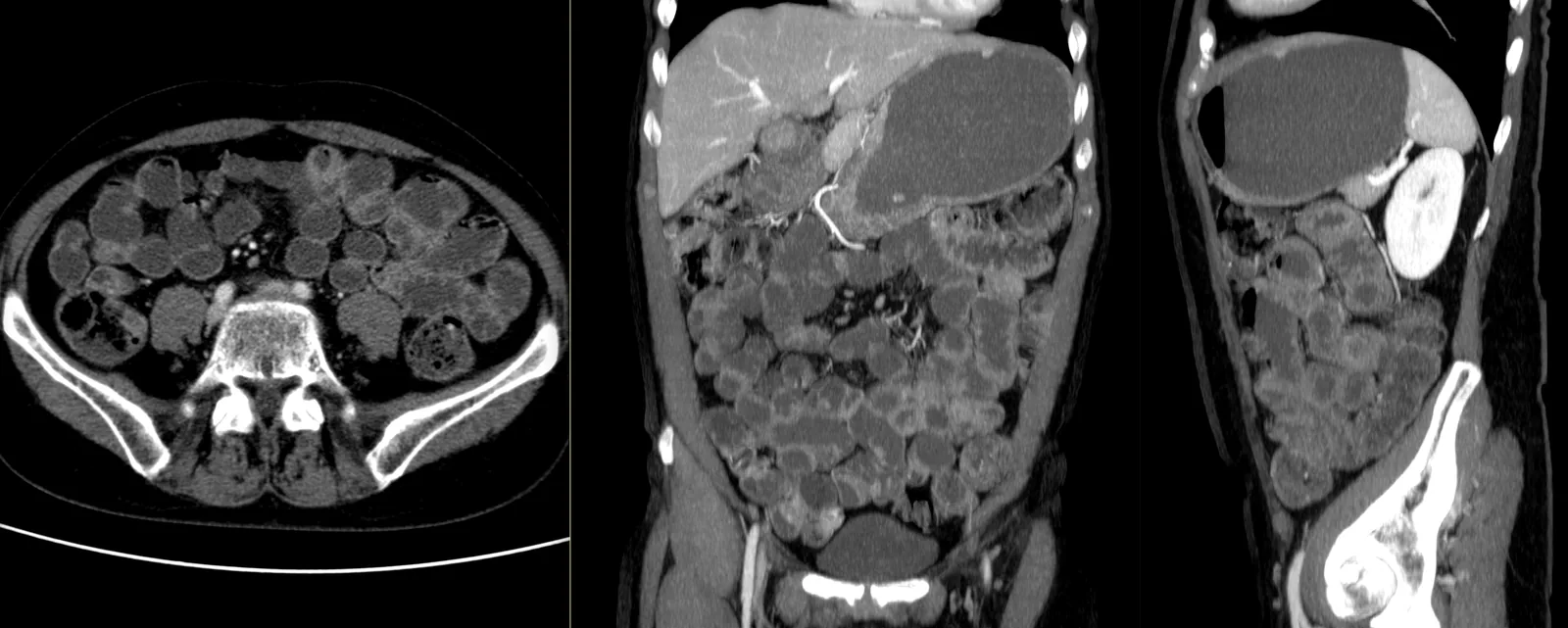
A CT enterography allows assessment of the small bowel — a part of the digestive tract that is difficult to study by other methods — including the wall of the bowel loops, their distension and the neighbouring structures (mesentery, lymph nodes, vessels). Distension with oral contrast is what makes the bowel wall clearly assessable; the intravenous contrast complements the study. It is often requested in the study of Crohn's disease — activity, extent and complications.
There is also MR enterography, the equivalent exam by magnetic resonance imaging, without radiation, often preferred in young patients who need repeated exams. The choice rests with the referring doctor. The report is produced by a radiologist; the clinical interpretation rests with your doctor.
When it is requested
- Crohn's disease — assessment of activity, extent and complications.
- Suspected inflammatory disease of the small bowel.
- Abdominal pain or chronic diarrhoea under investigation, according to clinical guidance.
- Gastrointestinal bleeding of unexplained cause, after endoscopies.
- Study of small-bowel changes identified on other exams.
- Assessment after bowel surgery, according to guidance.
What the exam is like
- 1Before
Before the day of the exam
Buy the product indicated when booking from the pharmacy — one 200 to 250 mL bottle of a lactulose solution — and bring it with you. Do not eat for 4 to 6 hours before the exam; you may drink water and, if you wish, a cup of tea up to 2 hours before. If you have recent blood tests with kidney function (last 3 to 6 months), it's advisable to bring them. Arrive 1 hour before your appointment time.
- 2
Preparing the oral contrast
At CRMA, the bottle you brought is mixed with 1.5 litres of water, forming the solution you will drink.
- 345 min
Drinking the solution
For the first 15 minutes, drink as quickly as you can; for the remaining time, continue at a normal pace, up to at least 1250 mL. The team supports you throughout the whole process.
- 4
Start of the exam
About 45 to 50 minutes after you start drinking, you go through to the CT room. You remove only the metal objects from the abdomen area (belts, fasteners, piercings).
- 510–20 min
Medication and intravenous contrast
A venous line is placed; a medicine that temporarily reduces the movements of the bowel may be given (it improves image quality), along with the intravenous iodinated contrast — it is normal to feel a passing warmth and a metallic taste. You will be asked to hold your breath for a few seconds during each acquisition.
- 6
Finishing
You can resume your normal day and your usual diet; we recommend drinking water throughout the day. The oral contrast may have a mild, passing laxative effect. If you notice passing blurred vision after the medication, wait for it to pass before driving.
Preparation
- Buy from the pharmacy: the product indicated when booking — one 200 to 250 mL bottle of a lactulose solution — and bring it on the day of the exam. The mixture is prepared at CRMA.
- Fasting: do not eat for 4 to 6 hours before the exam. You may drink water and, if you wish, a cup of tea up to 2 hours before.
- Arrival: 1 hour before your appointment time, for the preparation with the oral contrast. Allow 1h to 1h30 in total at the clinic.
- Kidney function: if you have recent blood tests (last 3 to 6 months) with creatinine/kidney function, it's advisable to bring them. They are not mandatory.
- Medication: keep your regular medication — including your diabetes and blood pressure medication, and metformin — unless your doctor advises otherwise.
- Iodinated contrast allergy: if you have had a reaction to iodinated contrast, let us know when booking. Each case is assessed individually by the radiologist.
- Metal objects: remove only those from the area being studied — belts, fasteners, piercings.
- Pregnancy: CT uses X-rays. If you are or think you may be pregnant, always let us know before the exam.
- Breastfeeding: always let us know. If the exam is with intravenous contrast, breastfeeding may continue as normal after the contrast is given (ACR/ESUR recommendation).
- After the exam: you can resume your usual diet. The oral contrast may have a mild, passing laxative effect. If you notice passing blurred vision after the medication, wait for it to pass before driving.
- Documents: referral, insurer/scheme card (if applicable), previous exams (endoscopies/CT/MRI) and recent blood tests, if you have them.
Results
The report is produced by a radiologist from the images. The report is available as a PDF (by email or printed) and the images on CD and on CRMA's imaging platform, to take to your doctor.