Mon–Fri 08:00–19:00 · Sat 08:00–13:00· Faro· +351 289 892 100· geral@crmalgarve.pt PTENFR
CRMA
Magnetic Resonance Imaging · By area

Magnetic Resonance Imaging of the Foot and Ankle

Allows highly detailed assessment of the ligaments, tendons, plantar fascia, cartilage and bones. No radiation. Often requested for sprains, tendinopathies, plantar fasciitis or chronic foot pain.

Duration20–30 min
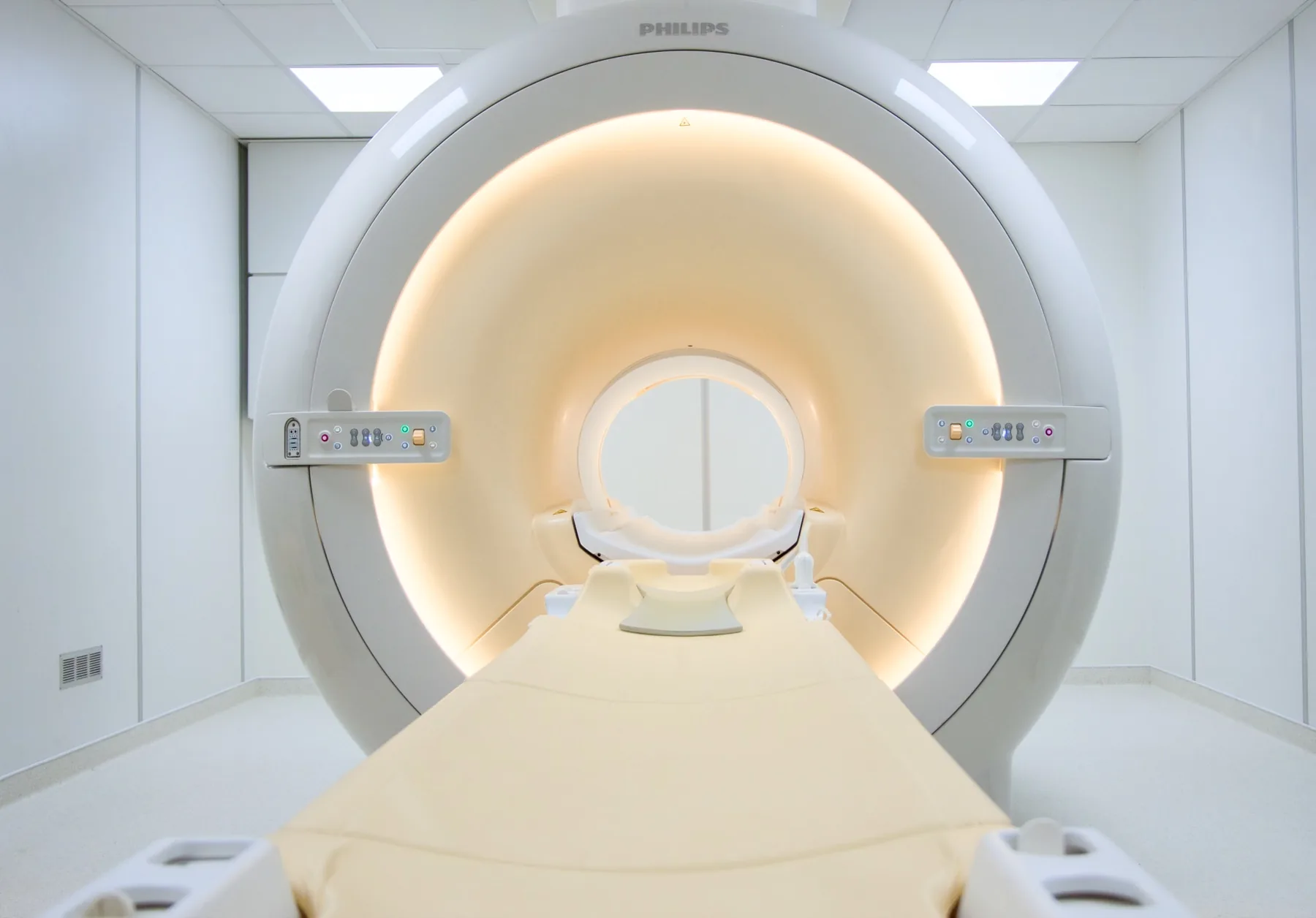
EquipmentPhilips 1.5T
ContrastRarely
PreparationNo fasting
CRMA's Magnetic Resonance Imaging (MRI) scanner, in Faro
In summary

A foot and ankle magnetic resonance (MRI) scan uses a magnetic field, with no radiation, to study in detail the ligaments, the tendons (including the Achilles tendon), the plantar fascia, the cartilage and the bones. It is often requested for sprains, tendinopathies, plantar fasciitis, stress fractures or chronic foot pain. At CRMA, in Faro, it takes about 20 to 30 minutes, you go in feet first (your head stays outside the tunnel) and, in most cases, it needs no contrast.

What is assessed in a foot and ankle MRI?

A foot and ankle MRI allows assessment of the ligaments (lateral ankle ligaments, deltoid ligament, syndesmosis), the tendons (Achilles, tibialis posterior, peroneal, flexor and extensor tendons), the plantar fascia, the cartilage, the bones (talus, calcaneus, malleoli, metatarsals) and the soft tissues, including looking for a Morton's neuroma.

Because it uses a magnetic field rather than X-rays, MRI involves no ionising radiation and gives great detail of the soft tissues and of bone injuries that are not visible on a plain X-ray. The report is written by a radiologist; the clinical reading is your treating doctor's.
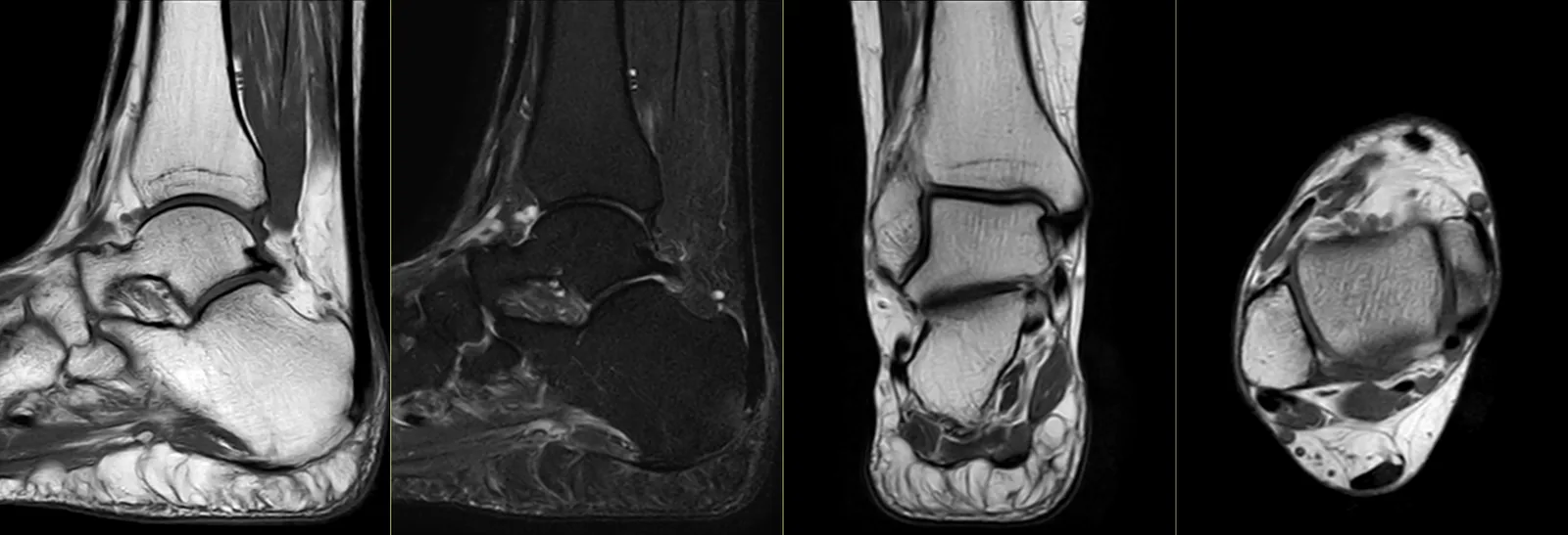
Foot and ankle magnetic resonance imaging: sagittal, coronal and axial slices showing the ligaments, the tendons, the cartilage and the bony structures.

When it is requested

  • Ankle sprain with persistent pain or a feeling of instability.
  • Suspected Achilles tendon injury (tendinopathy or rupture).
  • Tibialis posterior tendon dysfunction (adult acquired flatfoot).
  • Plantar fasciitis that does not improve or where the diagnosis is unclear.
  • Suspected stress fracture or a bone injury that does not show on an X-ray.
  • Osteochondral injury of the talus (cartilage + bone).
  • Morton's neuroma or other causes of forefoot pain.
  • Assessment before or after surgery to the foot or ankle.

What the exam is like

  • 1Before

    Reception and safety questionnaire

    Arrive 15 minutes early and fill in the safety questionnaire, if you haven't already done so online.

  • 2

    Changing room

    You undress and stay in underwear with no metal parts; for a foot study, you may need to take off your shoes and socks. You are given a gown.

  • 3

    Positioning

    The foot or ankle is placed inside a dedicated coil and you go into the scanner feet first.

  • 4

    Inside the scanner

    Because you go in feet first, most of your body and your head stay outside the tunnel — one of the best tolerated MRI exams for people with claustrophobia.

  • 520–30 min

    Image acquisition

    Several sequences with a characteristic noise; you are given ear protection. Contrast is rarely needed.

  • 6

    Finishing up

    You get dressed and can leave without needing anyone to accompany you.

Preparation

  • Fasting: not needed in general.
  • Changing room and clothing: as a rule, you undress and stay in underwear with no metal parts. For a foot/ankle study, you may need to take off your shoes and socks; the radiographer will tell you what to keep on. You are given a gown.
  • Devices and implants: note any pacemaker, neurostimulator and osteosynthesis hardware in the foot/ankle on the safety questionnaire.
  • Documents: your referral, your insurer or subsystem card (if using an agreement), and any previous exams.
  • Claustrophobia: you go in feet first and your head stays outside the tunnel — a well tolerated exam.

Results

The report is written by a radiologist from the images. The report is available as a PDF (by email or printed) and the images on CD and on CRMA's imaging platform, to take to your treating doctor — orthopaedic surgeon, physiatrist, family doctor or other.

Frequently asked questions

About foot and ankle MRI.

Does a foot/ankle MRI hurt?

No. It is a painless exam. There may be some discomfort from keeping the foot still for 20 to 30 minutes; the radiographer helps you find the most comfortable position.

How long does it take?

Generally between 20 and 30 minutes, which may increase with additional sequences.

I have claustrophobia. Can I have this exam?

Yes, generally with ease. You go in feet first, so most of your body and your head stay outside the tunnel. It is one of the best tolerated MRI exams for people with claustrophobia.

Does it study the foot or the ankle?

It depends on the doctor's request. The exam is directed at the area being studied — ankle, hindfoot, midfoot or forefoot. This is confirmed at booking.

Do I need contrast?

In most cases, no. Contrast is reserved for specific situations, such as suspected infection, a tumour, or assessment after surgery.

What is the difference between MRI and ultrasound of the foot?

Ultrasound assesses some tendons well and dynamically, and is quick; MRI gives a global, detailed view, including the bone and cartilage. They are complementary.

Can I have an MRI if I am pregnant?

Always let us know about confirmed or possible pregnancy. MRI does not use ionising radiation and may be performed at any stage of pregnancy when the doctor considers the information necessary; in the first trimester, as a precaution, the indication is weighed case by case. Contrast (gadolinium) is avoided throughout pregnancy unless clinically necessary.

Can I have it with hardware from foot or ankle surgery?

Many modern materials are MRI-compatible and may produce artefacts near the implant. Always note the hardware on the safety questionnaire and bring the documentation, if you have it.

Ready to book?

Book your foot or ankle MRI at CRMA.

In Faro, with personal service in PT, EN and FR. We'll confirm your booking with you during working hours.