
What is interventional radiology?
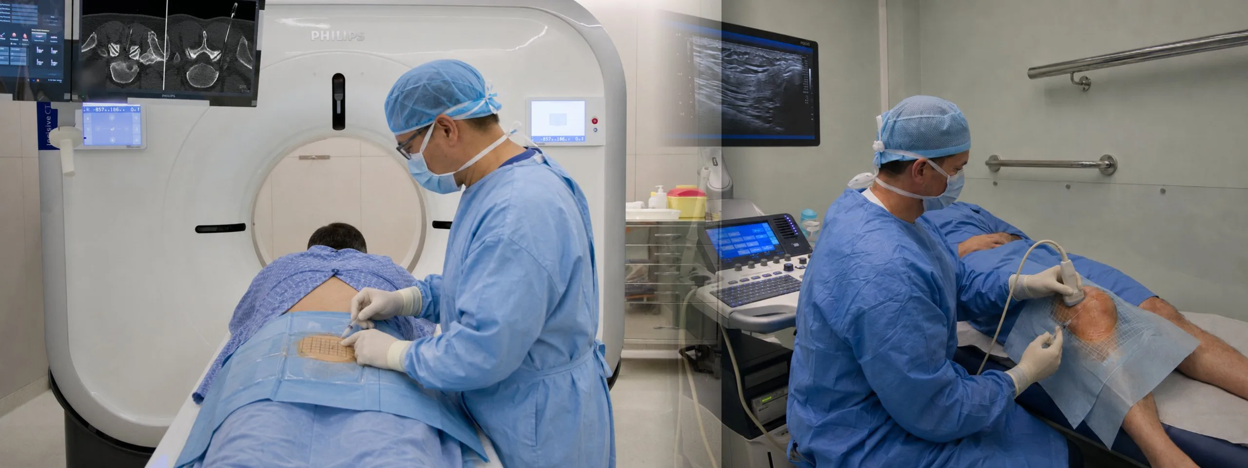
Interventional radiology uses imaging techniques — at CRMA, CT and ultrasound — to guide small therapeutic procedures with great precision. Instead of being used only to look, the imaging is used to treat: the radiologist follows the path of the needle in real time to the exact spot where the treatment needs to act.
These procedures are used mainly for the management of pain — for example, low back pain arising from the spine, joint pain (shoulder, hip, knee) or pain associated with tendons and bursae. The aim is to deliver the medication — usually an anti-inflammatory (corticosteroid) and a local anaesthetic, which may vary according to the patient's clinical condition and the radiologist's assessment — directly to the structure responsible for the pain, allowing small, targeted doses.
These are minimally invasive procedures: they are carried out with fine needles, through the skin, with no need for hospital admission. The response to treatment varies from person to person and is reviewed together with the doctor who follows your case.
CT-guided lumbar spine injections
CT allows the vertebrae and the surrounding structures to be seen in great detail, guiding the needle with millimetre precision — particularly important in the spine. At present, CRMA carries out these procedures in the lumbar and lumbosacral spine.
- Epidural (interlaminar) injection — medication delivered into the epidural space, around the membranes that surround the nerves. Common in low back pain that radiates down the leg.
- Periradicular (foraminal) injection — directed at the nerve root identified as the likely source of the pain, where it leaves the spine. Common in sciatica.
- Facet-joint injection — directed at the small posterior joints of the spine (zygapophysial joints), which can be a source of low back pain, particularly associated with osteoarthritis.
- Sacroiliac injection — directed at the sacroiliac joint, between the sacrum and the pelvis, a possible source of low back and buttock pain.
About radiation: CT guidance uses X-rays in low, localised doses, limited to what is needed to guide the procedure safely.
Ultrasound-guided injections and drainages
Ultrasound guides the needle in real time, with no radiation, and is particularly well suited to joints, tendons and soft tissues. At CRMA, these procedures are carried out with a modern ultrasound scanner.
- Joint injections — medication delivered inside joints such as the shoulder, the hip or the knee, usually in the context of persistent joint pain.
- Soft-tissue injections — directed at tendons and tendon sheaths, bursae (for example, bursitis) and other soft tissues.
- Ultrasound-guided drainages — draining fluid collections (for example, cysts or joint effusions) under ultrasound guidance.
What happens during a procedure
- 1Before
Contact and prior assessment
It all starts with your contact — through the form on this page or by phone. The case is assessed beforehand by the radiologist — by phone, in person, or based on your clinical documents — who confirms that the procedure is suitable and gives the preparation instructions.
- 2
Reception on the day of the procedure
Arrive with the lead time given at booking, fasting and with someone to accompany you. We confirm your regular medication and any allergies with you.
- 3
Positioning
You are positioned on the CT table or the ultrasound couch — usually lying face down for spinal injections.
- 4
Locating the point and cleaning the skin
The doctor identifies the entry point with the help of the imaging and cleans the skin. A local anaesthetic is given at the puncture site.
- 515–30 min
Guiding the needle with imaging
Under image control — CT or ultrasound — the needle is guided to the structure to be treated. You may feel pressure or brief discomfort; the local anaesthetic reduces the pain of the puncture.
- 6
Confirming the position and giving the treatment
In CT-guided procedures, the needle position is confirmed with a small amount of contrast. Once the position is confirmed, the medication is given and a simple dressing is applied.
- 7
Monitoring and going home
You stay under observation at the clinic for a short period. You go home with the person accompanying you — you may not drive after the procedure.
Preparation and safety
- Prior assessment: mandatory — carried out by the radiologist before booking (by phone, in person or from your documents).
- Anticoagulants / antiplatelet medicines: tell us about all the medication you take. Any stopping or adjustment is decided at the prior assessment, together with your treating doctor — never stop the medication on your own initiative.
- Allergies: tell us about allergies to local anaesthetics, anti-inflammatories/corticosteroids, iodinated contrast or adhesive dressings.
- Fasting: yes, it is required. The specific instructions are given at the time of booking.
- Diabetes: tell us if you are diabetic — the corticosteroid given can temporarily raise blood sugar.
- Pregnancy: tell us if you are or may be pregnant — particularly for CT-guided procedures.
- Active infection: a fever or infection on the day of the procedure should be reported — it may mean postponing.
- Someone to accompany you: please come with someone. After the procedure you may not drive.
- Documents: any previous imaging of the area to be treated (MRI, CT, X-rays) and a list of your regular medication.
- Reduced mobility: the clinic is accessible — a ramp with a lift and an adapted toilet. Let us know when you contact us.
After the procedure
In the first few hours you may feel numbness or a temporary reduction in strength in the treated area, as an effect of the local anaesthetic — this is expected and passes within a few hours. Over the next 24 to 48 hours the discomfort at the site may temporarily worsen before the effect of the medication is felt, as it sets in gradually. We recommend relative rest on the day itself, returning to your usual activities gradually.
The response to treatment varies from person to person. Assessing the result and deciding the next steps are done with the doctor who follows your case. You will receive from CRMA the record of the procedure carried out, which you can share with your doctor.
Warning signs
Contact us — or go to an emergency department — if in the following hours or days you develop a fever, intense and worsening pain, redness and warmth at the puncture site, or loss of strength or feeling that does not pass. These are rare situations, but they should be assessed without delay.