
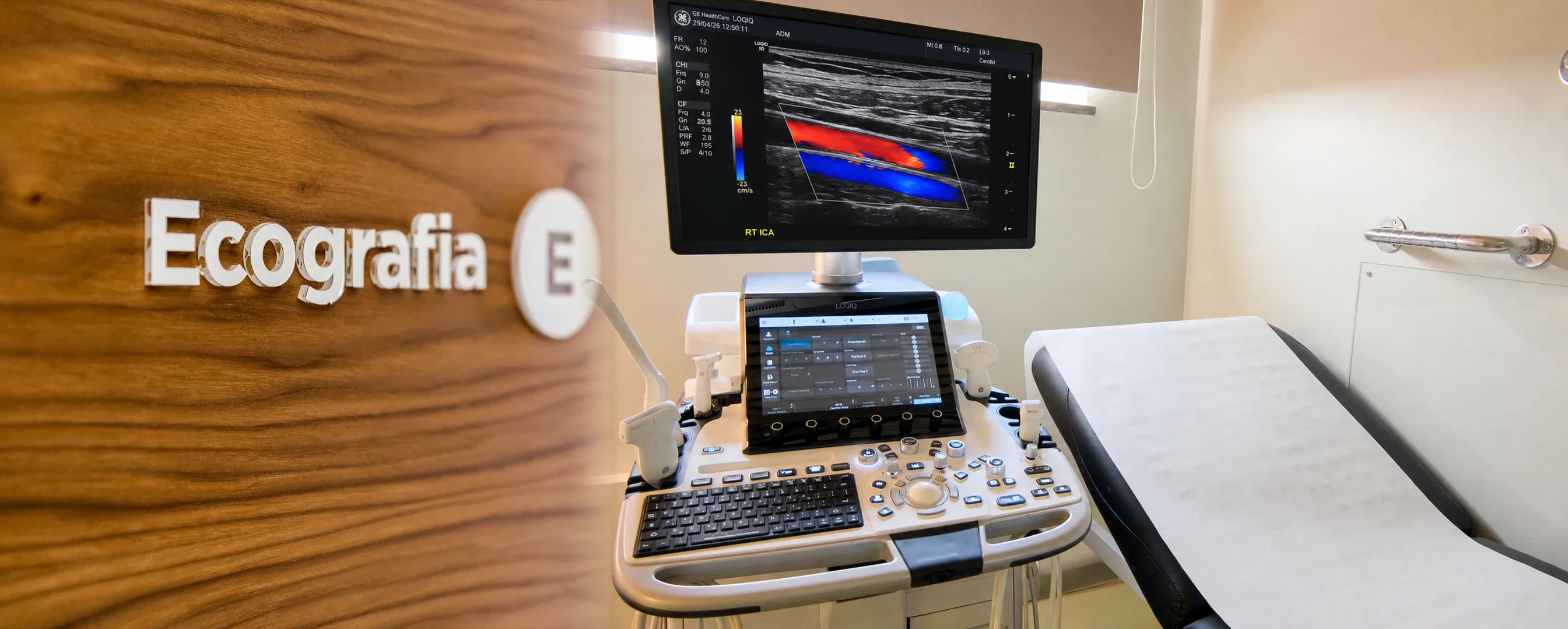
What can be assessed with a Doppler ultrasound?
A Doppler ultrasound adds the assessment of blood flow — direction, speed and obstacles to the circulation — to a standard ultrasound. At CRMA we perform, among others:
- Lower and upper limbs — arterial, venous or both: varicose veins and venous insufficiency, suspected thrombosis, peripheral arterial disease.
- Carotid and vertebral arteries — assessment of the arteries in the neck that supply the brain.
- Transcranial — intracranial circulation; can be combined with the carotid and ophthalmic study.
- Temporal arteries.
- Visceral — abdominal, renal, pelvic and uterine.
- Scrotal — on its own or as a complement to a scrotal ultrasound.
When it is requested
- Varicose veins, heavy or swollen legs — assessment of venous insufficiency.
- Suspected venous thrombosis.
- Carotid assessment — as directed by your treating doctor, for example after a carotid bruit or in the context of vascular risk.
- Leg pain when walking (claudication) — arterial study.
- Follow-up of already known or operated vascular conditions.
The exam is always requested by the referring doctor — CRMA also works with visiting specialists in vascular surgery and neurology.
What the exam is like
- 1Before
Reception
Arrive at the time given when booking, with your referral and any previous exams, if you have them.
- 2
Positioning
Lying down or standing, depending on the area to be studied — for a venous study of the limbs you may be asked to stand so that reflux can be assessed.
- 315–35 min
Gel and probe
The doctor applies gel and slides the probe along the vessels, recording images and the sounds of the flow. You may hear the characteristic «whoosh» of the blood circulating.
- 4
Finishing up
The gel is wiped off and you can carry on with your day with no restrictions.
Preparation
Most Doppler studies need no preparation. The usual exception, confirmed when you book:
- Abdominal, visceral or renal Doppler: fast for 4 to 6 hours, depending on the study and on the instructions given when booking, to reduce bowel gas.
- Clothing: comfortable and easy to adjust to the area being studied.
- Regular medication: continue as usual, unless your doctor tells you otherwise.
Results
The report is written by a radiologist. The report is available as a PDF (by email or printed) and the images on CRMA's imaging platform, to take to your treating doctor. With written authorisation, they can be sent directly to the referring doctor.