
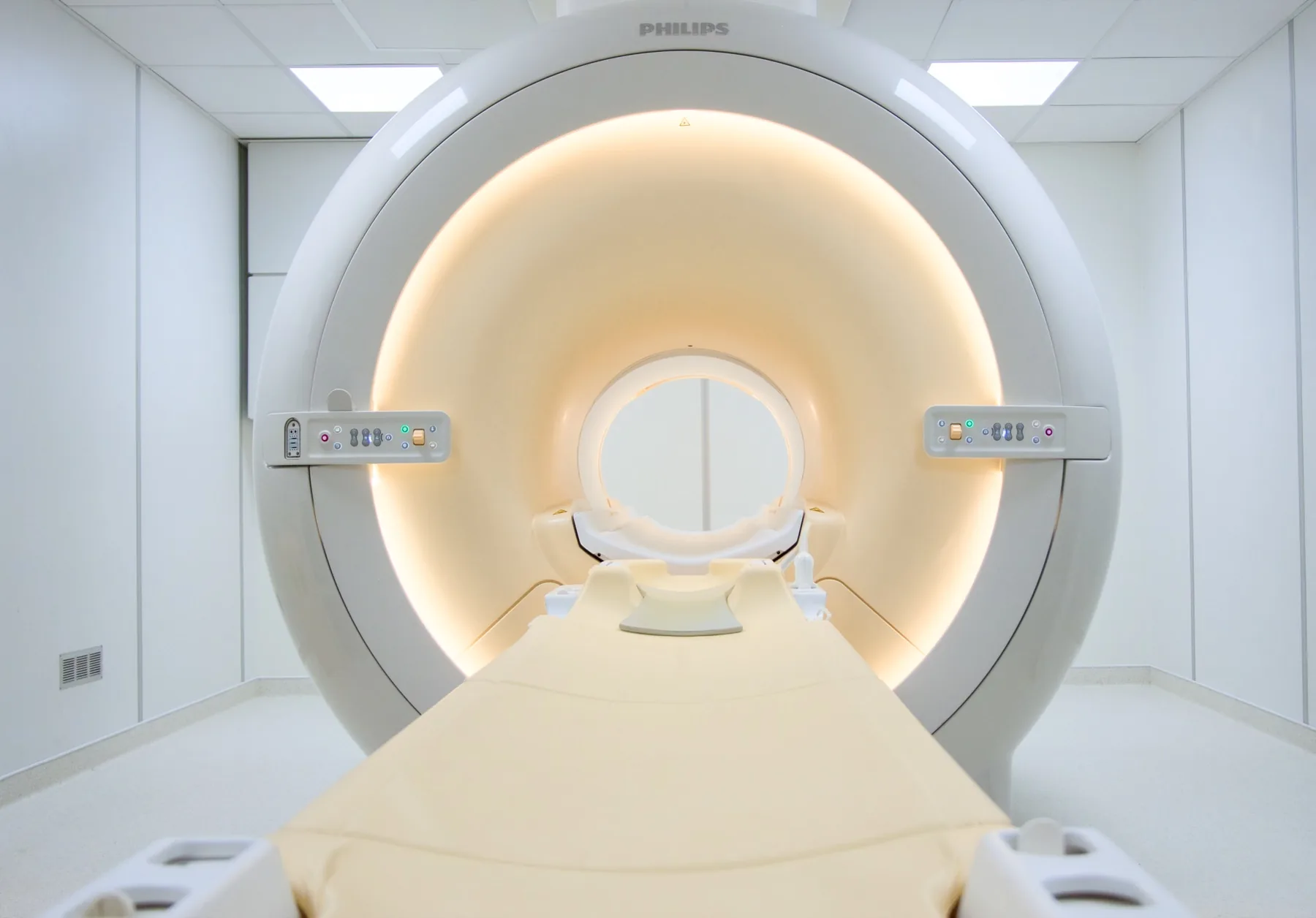
What is assessed in a prostate MRI?
Multiparametric prostate MRI allows assessment of the prostate gland — its different zones —, the seminal vesicles, the capsule and its relationship with the surrounding structures. "Multiparametric" means it combines several types of sequence (T2, diffusion and a dynamic contrast-enhanced study), which gives more complete information.
The result helps the doctor decide the next steps (for example, whether and where to perform a biopsy). Because it uses a magnetic field, it involves no ionising radiation. The report is written by a radiologist; the clinical decision rests with the urologist or treating doctor.
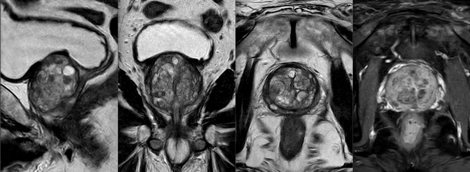
When it is requested
- Raised PSA and/or an abnormal finding on digital rectal examination, before deciding on a biopsy.
- Biopsy guidance — to direct the sampling to suspicious areas (targeted or fusion biopsy).
- Active surveillance of a known low-risk prostate cancer.
- Local staging of a confirmed cancer, before a treatment decision.
- Suspected recurrence after treatment.
What the exam is like
- 1Before
Reception and safety questionnaire
Arrive 15 minutes early and fill in the safety questionnaire. Note a recent kidney-function blood test, if you have one.
- 2
Preparation
Use a cleansing rectal enema (Microlax® or Cliss-Go®) at home — on waking in the morning or around lunchtime, depending on the time of your exam. No abstinence is needed.
- 3
Positioning
Empty your bladder immediately beforehand. You lie on your back; the study uses a surface coil (with an element built into the table) — no endorectal coil.
- 4
Inside the scanner
The pelvic region sits at the centre of the magnet, so part of your body goes into the tunnel. You stay in contact with the radiographer.
- 530–45 min
Multiparametric acquisition
Several sequences, including diffusion and a dynamic contrast-enhanced study.
- 6
Finishing up
The intravenous line is removed; you get dressed and can leave without needing anyone to accompany you.
Preparation
- Changing room and clothing: as a rule, you undress and stay in underwear with no metal parts — usually underpants and socks. Depending on the area, the radiographer may allow you to keep some clothing on. You are given a gown.
- Cleansing rectal enema: at home before you come — Microlax® or Cliss-Go®, on waking in the morning or around lunchtime depending on the time of your exam.
- Ejaculatory abstinence: not needed.
- Bladder: empty your bladder immediately before going in.
- After a biopsy: no specific interval is required; please tell us, even so, that you have had one.
- Contrast and kidney function: a recent kidney-function blood test may be requested.
- Documents: your referral, your PSA value and any previous reports (ultrasound, biopsy, previous MRI).
Results
The report is written by a radiologist. The report is available as a PDF (by email or printed) and the images on CD and on CRMA's imaging platform, to take to your urologist or treating doctor.