
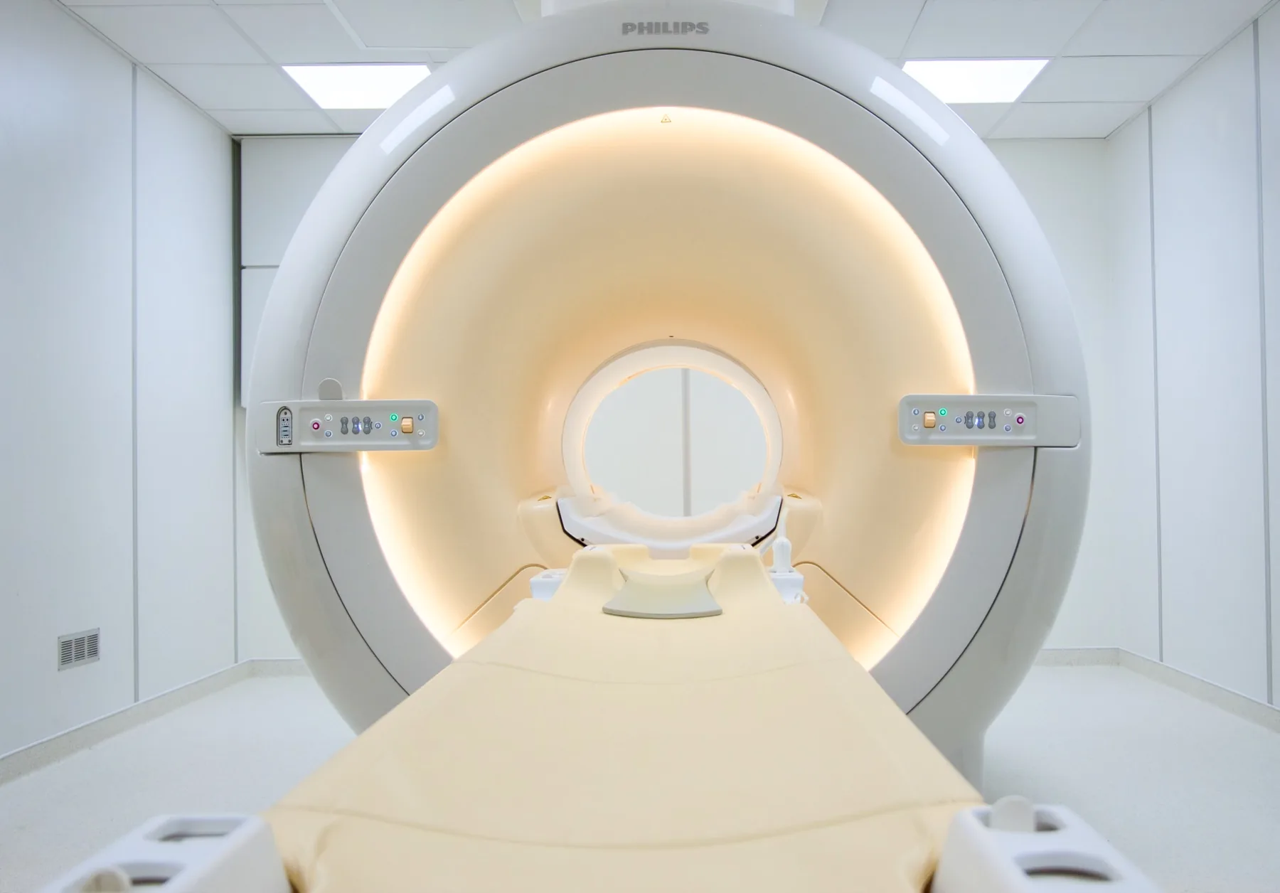
What is assessed in a head MRI?
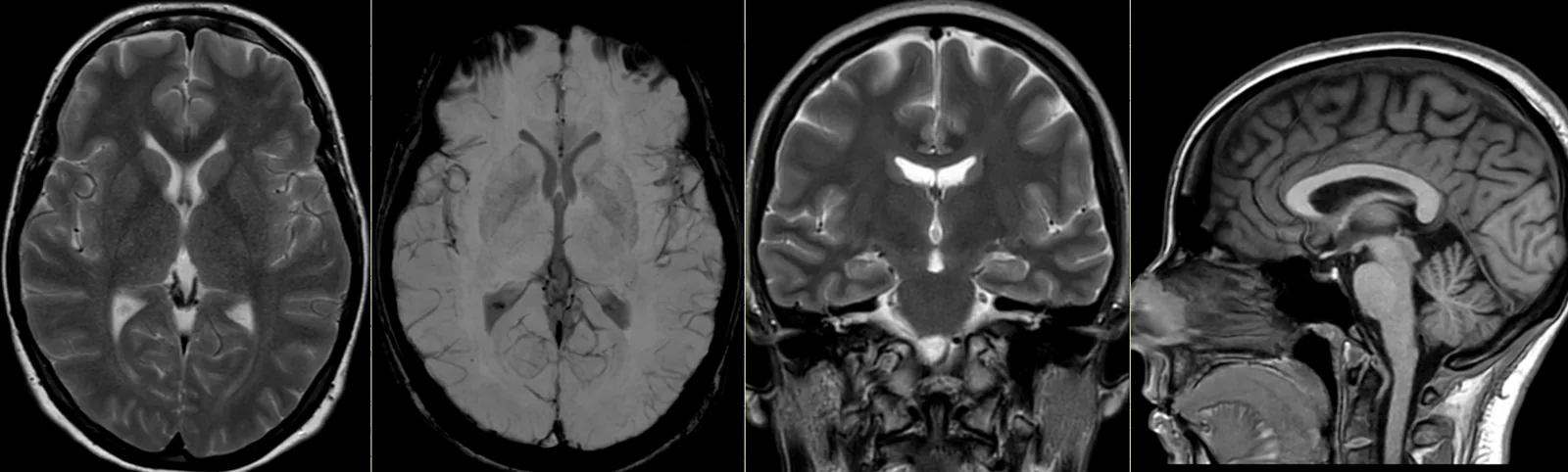
Head MRI allows assessment of the brain — the cerebrum, the cerebellum and the brainstem —, distinguishing white matter from grey matter, the ventricles and the cerebrospinal fluid spaces, the cranial nerves and structures such as the pituitary gland or the orbits (in targeted studies). With MR angiography, it also studies the vessels of the brain, without radiation.
MRI is particularly sensitive in the study of the nervous-system tissues. Diffusion sequences are very useful in the early assessment of ischaemic stroke. Because it uses a magnetic field, it involves no ionising radiation. The report is written by a radiologist; the clinical interpretation rests with your treating doctor.
When it is requested
- Persistent headaches, severe or with atypical features.
- Dizziness, vertigo or balance problems.
- Suspected stroke — MRI diffusion is very sensitive, above all in ischaemic stroke.
- Epilepsy or seizures.
- Changes in vision or hearing (tinnitus, sensorineural hearing loss).
- Demyelinating conditions (e.g. multiple sclerosis), often with a study of the cervical spinal cord.
- Suspected space-occupying lesion and its follow-up.
- Memory changes or study of the pituitary gland.
What the exam is like
- 1Before
Reception and safety questionnaire
Arrive 15 minutes early and fill in the safety questionnaire, if you haven't already done so online.
- 2
Changing room
Remove any metal objects; you may be asked to remove eye make-up and contact lenses, as some products contain metallic particles.
- 3
Positioning
Your head is supported and surrounded by a dedicated coil (like an open helmet), with cushions to keep it still.
- 4
Inside the scanner
Your head is inside the tunnel — this is the MRI exam where the sense of an enclosed space is most noticeable. You stay in contact with the radiographer and are given an emergency call button.
- 520–40 min
Image acquisition
Several sequences with a characteristic knocking noise; you are given ear protection. When indicated, contrast is administered (tumour, infection, demyelinating disease).
- 6
Finishing up
You get dressed and can leave without needing anyone to accompany you, unless told otherwise.
Preparation
- Changing and clothing: as a rule, you undress and stay in underwear with no metal parts — usually pants and socks. Depending on the area, the radiographer may allow you to keep some clothing on. You are given a gown.
- Fasting: not needed in general, even when contrast is expected.
- Make-up and lenses: avoid eye make-up on the day of the exam (some contain metallic particles); you may be asked to remove contact lenses.
- Metal objects: remove hairpins, earrings, piercings, removable dentures and hearing aids.
- Devices and implants: note on the questionnaire any pacemaker, neurostimulator, cerebral aneurysm clips and cochlear implant — some are incompatible and require a prior assessment.
- Claustrophobia: your head is inside the tunnel. If you are anxious about this, contact us before the day of the exam.
- Children: sedation may be needed, arranged with the doctor. Let us know when booking.
Results
The report is written by a radiologist from the images. The report is available as a PDF (by email or printed) and the images on CD and on CRMA's imaging platform, to take to your treating doctor — neurologist, neurosurgeon, family doctor or other.