
Referrers.
A space for referring doctors. Here you'll find practical information on how to refer, how to access your patients' reports and images, and how to reach CRMA's clinical team directly, in Faro.
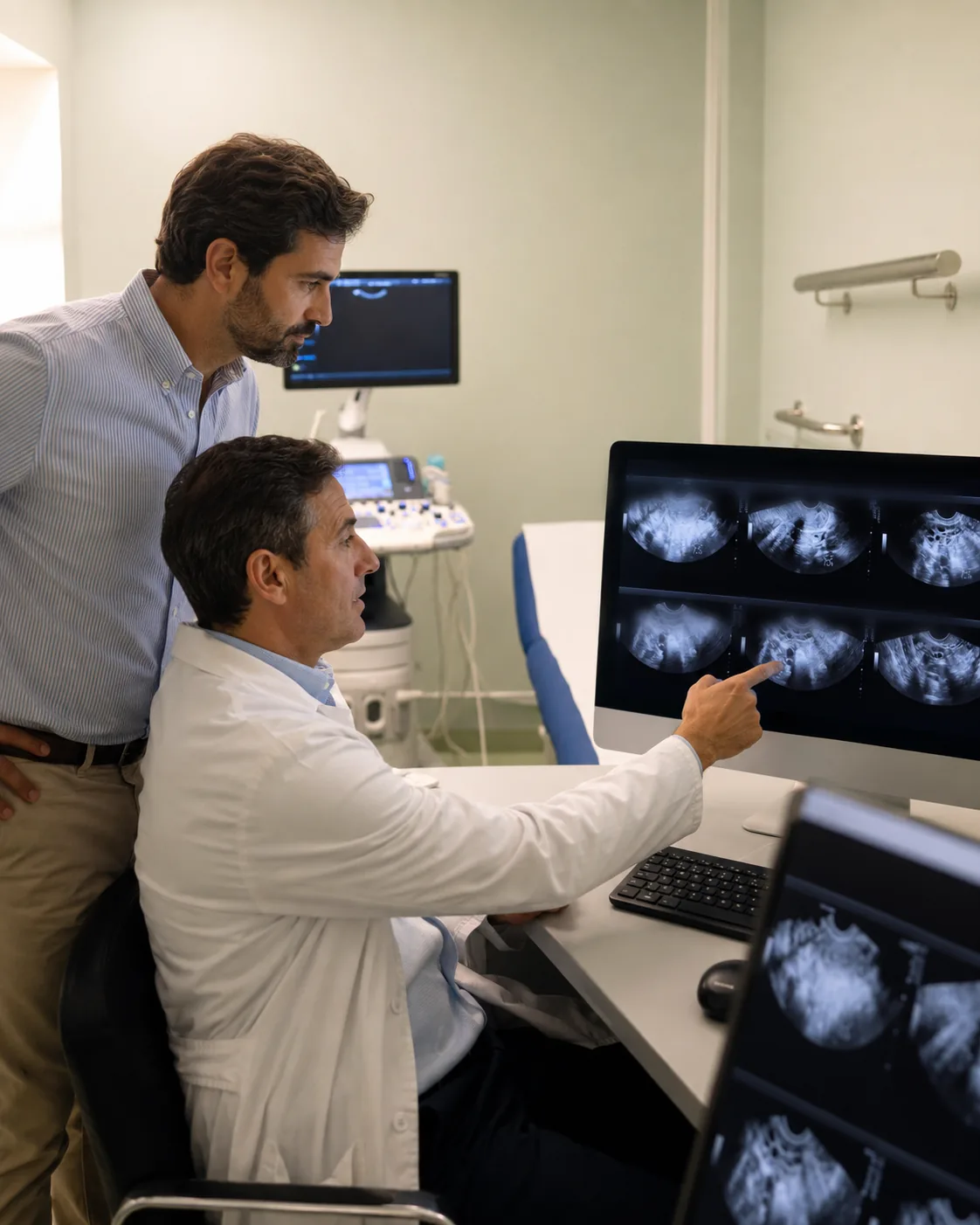
How to refer exams to CRMA.
CRMA accepts requests from any healthcare provider, in any usual format — paper, digital or electronic prescription (SNS 24 / paperless prescription). These good practices help the team prepare the most appropriate exam.
Region and clinical indication
In MRI and CT, the region to be studied (right knee, abdomen, brain) and the clinical reason (pain, suspected lesion, follow-up) allow the radiologist to define the most appropriate protocol.
Whether contrast is needed
In contrast-enhanced CT and MR angiography, the use of contrast depends on the type of exam and, in MRI, on the type of sequence, and is a clinical decision shared between the referrer and the radiologist responsible for the exam.
Known contraindications
Pacemakers, prostheses, contrast allergies, impaired kidney function, marked claustrophobia or pregnancy. When this information is already known, noting it on the request speeds up the process.
Referrer identification
A legible name, specialty, Ordem dos Médicos (Portuguese Medical Association) licence number and a direct contact. It is often necessary to contact the referrer for clinical clarification or to report relevant findings.
Sending the report to the referrer
By default, the report is given to the patient. If you would prefer to receive it directly — by secure email or post — please note this on the request, with the patient's consent.
CRMA performs CT-guided lumbar-spine injections (epidural/interlaminar, periradicular, facet joint and sacroiliac) and ultrasound-guided joint, soft-tissue and drainage procedures, for pain management. Booking is always preceded by a prior assessment by the radiologist. When referring, please include the relevant prior imaging and the patient's regular medication (in particular anticoagulants and antiplatelet agents). Contact: +351 289 892 100. See the modality page →
How to receive your patients' reports and images.
CRMA makes the report and images of each exam available. The current process offers three ways to receive them, according to the patient's and the referrer's preference.
Report by email (PDF)
The report is sent as a PDF to the patient's email, within the timeframe appropriate to the type of study. To receive a copy in parallel, simply note it on the request, with the patient's consent.
Images on CD (DICOM)
The images in DICOM format are available on CD, for collection at CRMA's reception. The CD includes a standalone image viewer, compatible with any computer.
Online imaging platform
For CT and MRI exams, the images are also accessible through CRMA's imaging platform (resultados.crmalgarve.pt), with credentials given to the patient — who can share them with their doctor.
Thirteen modalities across five areas.
CRMA operates the main modalities of medical imaging and functional testing, including image-guided procedures. The detailed list, with preparation and indications, is on the general exams page.
X-ray imaging
- Computed Tomography (CT) and CT angiography, including Cardiac + Calcium Score
- Mammography with 3D Tomosynthesis
- Bone Densitometry
- Conventional Radiology (X-ray)
- Digital Orthopantomogram
Magnetic resonance imaging
- Magnetic Resonance Imaging (MRI), MR angiography and 4D MRA, with no ionising radiation
Ultrasonography
- Ultrasound (general, breast, obstetric, gynaecological, soft tissue, musculoskeletal)
- Doppler (vascular, abdominal, transcranial, scrotal, ophthalmic)
Functional testing
- Cardiopulmonary Testing (Echocardiogram, ECG, Holter, ABPM, Spirometry)
- Electromyography (EMG)
- Electroencephalography (EEG)
- Laboratory Tests (in partnership with AQUALAB)
Image-guided procedures
- Interventional Radiology — CT-guided lumbar-spine injections; ultrasound-guided injections and drainages, for pain management
Common clinical indications by modality.
A summary of the situations in which each modality is usually appropriate, in line with international referral guidelines — RCR iRefer (Royal College of Radiologists) and ACR Appropriateness Criteria — and exam-specific standards (BI-RADS, PI-RADS, Fleischner criteria, ISCD, CAD-RADS). They do not replace the referrer's clinical judgement or the radiologist's assessment.
Supporting content, general in nature. If in doubt about the most appropriate modality, contact the team — the final decision is always shared between the referrer and the radiologist.
Magnetic Resonance Imaging (MRI) and MR angiography
No ionising radiation; high soft-tissue contrast resolution. Usually indicated to assess:
- Neurological: headaches with red-flag features, suspected ischaemia/stroke (diffusion sequences), demyelinating disease (multiple sclerosis), epilepsy, central nervous system tumours, and pituitary and cerebellopontine-angle pathology.
- Spine: radiculopathy or neurological deficit, suspected disc herniation, canal stenosis, myelopathy, spinal cord compression and spondylodiscitis.
- Musculoskeletal: meniscal and ligament injuries (knee), rotator cuff and labral pathology (shoulder), chondral lesions, bone oedema/occult fracture, osteonecrosis and soft-tissue lesions.
- Abdomen and pelvis: characterisation of liver lesions, MRCP (biliary tract), pancreatic and gynaecological pelvic pathology; multiparametric prostate MRI (PI-RADS) in suspected prostate cancer and its local staging.
Always confirm contraindications (implanted devices, metallic foreign bodies) through the safety questionnaire.
Computed Tomography (CT), CT angiography and Cardiac CT
Fast and widely available, with dose optimised to what is needed. Usually indicated to assess:
- Neurological and emergency: head trauma, suspected intracranial haemorrhage and acute stroke (to exclude haemorrhage).
- Chest: characterisation and follow-up of a pulmonary nodule (Fleischner criteria), oncological staging, suspected pulmonary thromboembolism (CT angiography) and interstitial lung disease (high resolution).
- Abdomen and pelvis: acute abdominal pain (appendicitis, diverticulitis, obstruction), urinary stones (Uro-CT), staging and characterisation of masses.
- Cardiovascular: coronary CT angiography in chest pain of low-to-intermediate probability; Calcium Score (Agatston) in cardiovascular risk stratification; CT angiography of the aorta, supra-aortic trunks and limbs.
- Musculoskeletal and head/neck: complex fractures and surgical planning, paranasal sinuses and dental scan (implantology).
Iodinated contrast when indicated — consider kidney function and any history of a contrast reaction.
Ultrasound and Doppler
No radiation; often first-line, especially in pregnant women and children. Usually indicated to assess:
- Abdomen and kidney: right-upper-quadrant pain, gallstones, steatosis, liver and kidney characterisation, abdominal aorta.
- Pelvis, gynaecology and obstetrics: pregnancy monitoring and uterine and adnexal pathology (abdominal or transvaginal route).
- Superficial: thyroid, breast (complementing mammography, especially in dense breasts), soft tissue, musculoskeletal (tendons, effusions) and scrotal.
- Doppler: suspected deep vein thrombosis and limb venous insufficiency, carotid stenosis (supra-aortic trunks), limb arterial study and renal/visceral Doppler.
Mammography with tomosynthesis
Usually indicated to assess:
- Screening for breast cancer in asymptomatic women, according to screening standards and individual risk.
- Diagnostic work-up of symptoms — a palpable lump, retraction, nipple discharge or skin changes.
- Complementary breast ultrasound in dense breasts and to clarify findings.
- Follow-up and structured classification (BI-RADS).
The starting age and frequency of screening follow the standards in force and risk assessment.
Bone densitometry (DEXA)
Quantifies bone mineral density (T-score / Z-score, per the ISCD). Usually indicated to assess:
- Post-menopausal women and men with osteoporosis risk factors.
- A history of a fragility fracture.
- Prolonged corticosteroid therapy or other causes of secondary osteoporosis.
- Fracture-risk estimation (FRAX) and monitoring of treatment response (interval per the ISCD).
X-ray (conventional radiology)
Low dose, fast and first-line for many complaints. Usually indicated to assess:
- Trauma: suspected fracture and initial bone assessment.
- Chest: respiratory symptoms, heart failure and selected pre-operative work-up.
- Spine: weight-bearing study, alignment and scoliosis.
- Musculoskeletal: initial assessment of osteoarthritis and joint pain.
Channels dedicated to referrers.
For clinical questions, protocol choice, report clarification or requests for additional copies, speak directly with the CRMA team.
By phone
+351 289 892 100 (cost of a call to a national landline). Identify yourself as a referring doctor when you call, for direct routing. Mon–Fri 08:00–19:00 · Sat 08:00–13:00.
By email
geral@crmalgarve.pt. For technical clinical questions, report clarification and requests for additional copies of reports or images.
Clinical Directorate
Dr. Pedro Ramos — radiologist, Clinical Director of CRMA (Ordem dos Médicos licence no. 28284). For institutional matters, partnerships or complex clinical cases, ask to be routed to the Clinical Directorate when you contact the clinic. For administrative matters, use the contact page.
Count on CRMA for your patients.
See the full list of exams or get in touch by phone (+351 289 892 100) or email. We confirm the booking with the patient during working hours.