
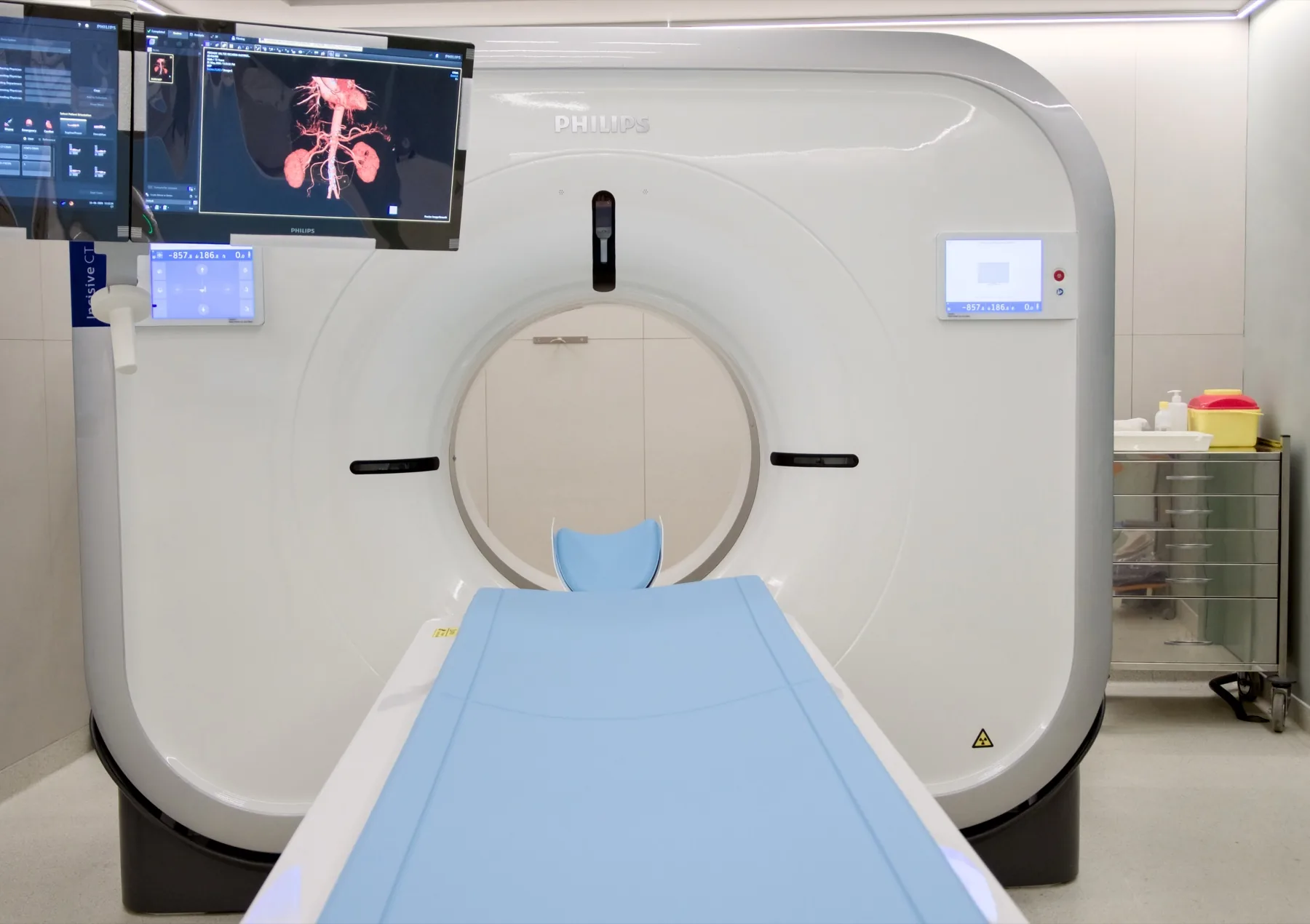
What is assessed in an abdomen and pelvis CT scan?
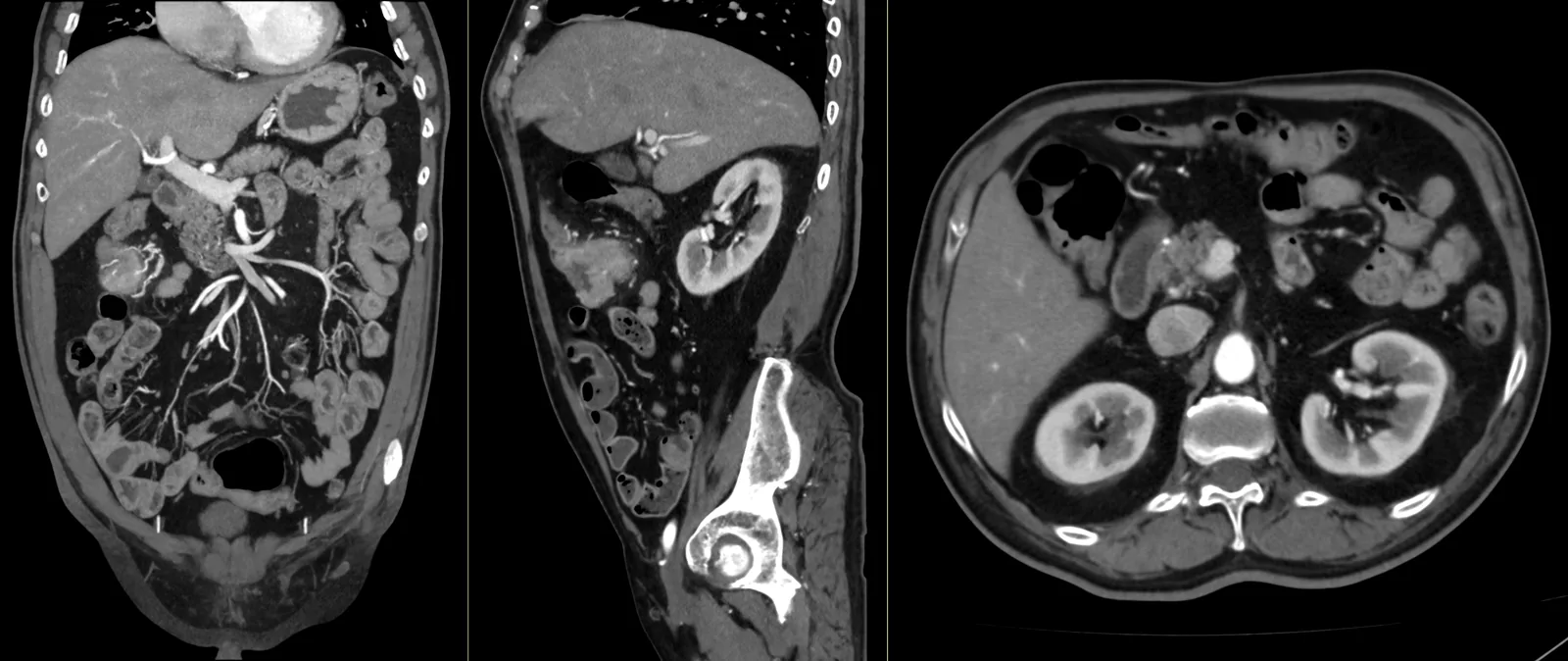
An abdomen and pelvis CT scan allows assessment of the liver, the gallbladder and bile ducts, the pancreas, the spleen, the kidneys and urinary tract, the adrenal glands, the digestive tract, the bladder and the pelvic organs, as well as the lymph nodes, the vessels and the bony structures of the region.
In most studies, intravenous iodinated contrast is given, which improves the characterisation of the organs and of any lesions. In some cases, depending on the clinical information, oral contrast is also given, to distend and outline the digestive tract. Targeted studies have their own protocols: the urinary tract is studied with Uro-CT and the small bowel with CT enterography. The report is produced by a radiologist; the clinical interpretation rests with your doctor.
When it is requested
- Abdominal pain under investigation, according to clinical guidance.
- Characterisation of ultrasound findings — lesions of the liver, the kidneys or other organs.
- Suspected inflammatory processes — for example, diverticulitis or appendicitis.
- Study of the bile ducts and the pancreas.
- Urinary stones — usually with a non-contrast protocol; the targeted study of the urinary tract is Uro-CT.
- Staging and follow-up in a cancer setting, according to your doctor's guidance.
- Assessment after abdominal or pelvic surgery.
- After abdominal injury, according to clinical guidance.
What the exam is like
- 1Before
Reception
Arrive 15 minutes early — at least 30 minutes early if the exam includes oral contrast (we let you know when booking). You do not need to fast, even with contrast; if you have recent blood tests with kidney function (last 3 to 6 months), it's advisable to bring them.
- 2
Oral contrast (in some cases)
Depending on the clinical information, you may be given an oral contrast to drink before the exam, to outline the digestive tract. In that case, you should arrive at least 30 minutes before your appointment — we let you know when booking.
- 3
Preparation
You remove only the metal objects from the abdomen and pelvis area: belts, fasteners, piercings. As a rule, you keep your own clothes on, as long as they have no metal components in the area.
- 4
Positioning
Lying on your back on the table, usually with your arms above your head.
- 510–20 min
Image acquisition
The table moves through the scanner's open ring. You will be asked to hold your breath for a few seconds during each acquisition. With intravenous contrast, it is normal to feel a passing warmth — sometimes with a feeling of needing to urinate — and a metallic taste; these pass within a few minutes.
- 6
Finishing
You can leave, drive and resume your normal day. We recommend drinking water throughout the day.
Preparation
- Fasting: not required, even with contrast — which is usual for this study. Have a light meal before you come and drink water freely up to the time of your exam. If the exam includes oral contrast, you stop eating when you start drinking the solution at the clinic — the team will tell you at what time.
- Kidney function: if the exam is with contrast and you have recent blood tests (last 3 to 6 months) with creatinine/kidney function, it's advisable to bring them. They are not mandatory.
- Medication: keep your regular medication — including your diabetes and blood pressure medication, and metformin — unless your doctor advises otherwise.
- Iodinated contrast allergy: if you have had a reaction to iodinated contrast, let us know when booking. Each case is assessed individually by the radiologist.
- Oral contrast: when indicated by the clinical information, it is provided at CRMA before the exam — arrive at least 30 minutes before your appointment.
- Metal objects: remove only those from the area being studied — belts, fasteners, piercings.
- Clothing: as a rule, you keep your own clothes on, as long as they have no metal components in the area.
- Breathing: during the exam you will be asked to hold your breath for a few seconds — the radiographer guides you at the time.
- Pregnancy: CT uses X-rays. If you are or think you may be pregnant, always let us know before the exam.
- Breastfeeding: always let us know. If the exam is with contrast, breastfeeding may continue as normal after the contrast is given (ACR/ESUR recommendation).
- Documents: referral, insurer/scheme card (if applicable), previous exams (ultrasound/CT/MRI) and recent blood tests, if you have them.
Results
The report is produced by a radiologist from the images. The report is available as a PDF (by email or printed) and the images on CD and on CRMA's imaging platform, to take to your doctor.