
What is assessed in a sinus CT scan?
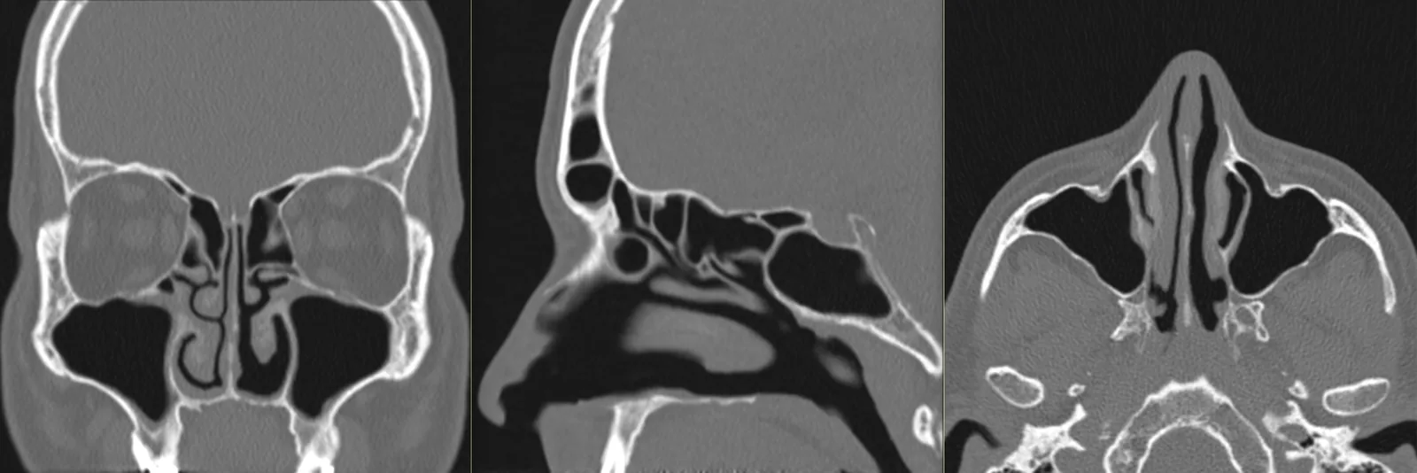
A sinus CT scan allows assessment of the four pairs of paranasal sinuses — maxillary, ethmoid, frontal and sphenoid —, the nasal cavities, the nasal septum and the drainage structures (the ostiomeatal complex), as well as the surrounding bony walls. It is the reference imaging exam for the anatomical study of this region.
It allows characterisation of mucosal thickening, filling of the sinuses (secretions, polyps), anatomical variants that predispose to obstruction and post-surgical changes — information that is useful to the ENT specialist, particularly in planning endoscopic sinus surgery. The report is produced by a radiologist; the clinical interpretation rests with your doctor.
When it is requested
- Chronic or recurrent sinusitis — characterisation of the extent and of the causes of obstruction.
- Persistent nasal obstruction — deviated septum, anatomical variants, polyps.
- Planning endoscopic sinus surgery (FESS) — an anatomical map for the surgeon.
- Assessment after surgery of the paranasal sinuses.
- Headache in the region of the face, according to clinical guidance.
- Changes in the sense of smell, according to clinical guidance.
- Targeted study of the maxillary sinuses — including in the context of disease of dental origin.
What the exam is like
- 1Before
Reception
Arrive 15 minutes early, with your referral and previous exams, if you have them.
- 2
Preparation
You remove only the metal objects from the head area: hairpins, earrings, piercings, glasses, removable dentures. As a rule, you keep your own clothes on.
- 3
Positioning
Lying on your back on the table, with your head in a dedicated support.
- 45–10 min
Image acquisition
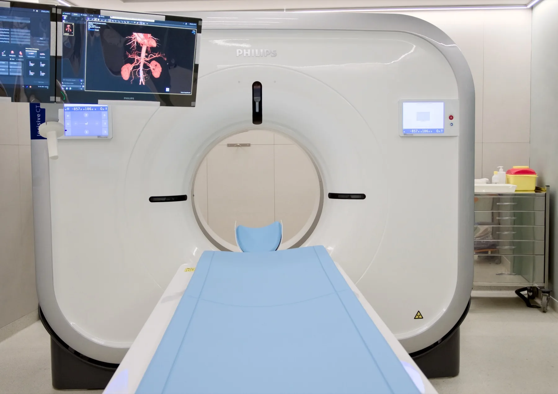
The table moves through the scanner's open ring. You just need to keep your head still for a few brief moments — acquisition takes seconds.
- 5
Finishing
You can leave, drive and resume your normal day straight away.
Preparation
- Fasting: not needed.
- Contrast: rarely needed. Even when it is used, fasting is not required; only the kidney-function recommendations apply (recent blood tests from 3 to 6 months, if you have them) — we let you know when booking.
- Medication: keep your regular medication.
- Metal objects: remove only those from the area being studied — hairpins, earrings, piercings, glasses, removable dentures.
- Clothing: as a rule, you keep your own clothes on.
- A cold on the day of the exam: an acute cold can temporarily change the appearance of the lining. If you have a heavy cold, talk to us — together with your doctor's guidance, we assess whether to keep the date.
- Pregnancy: CT uses X-rays. If you are or think you may be pregnant, always let us know before the exam.
- Children: let us know when booking, so we can prepare the exam and adjust the protocol.
- Documents: referral, insurer/scheme card (if applicable), previous exams (X-ray/CT).
Results
The report is produced by a radiologist from the images. The report is available as a PDF (by email or printed) and the images on CD and on CRMA's imaging platform, to take to your doctor.