
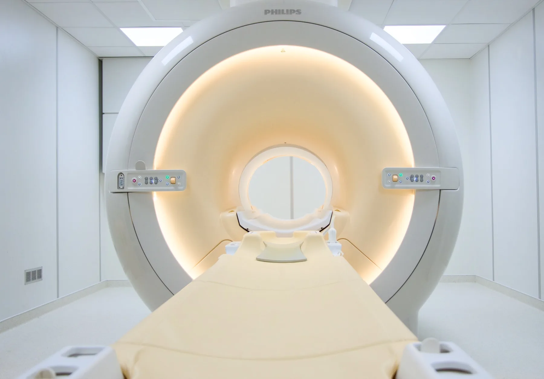
What is assessed in a hip MRI?
Hip MRI allows assessment of the structures of the hip joint — the femoral head and the acetabulum, the acetabular labrum, the cartilage, the capsule, the tendons (gluteal, iliopsoas), the bursae (trochanteric) and the bones of the pelvis and femur.
Because it uses a magnetic field rather than X-rays, MRI involves no ionising radiation and is particularly useful in situations that a plain X-ray does not show, such as early changes in the femoral head or soft-tissue injuries. The report is written by a radiologist; the clinical interpretation rests with your treating doctor.
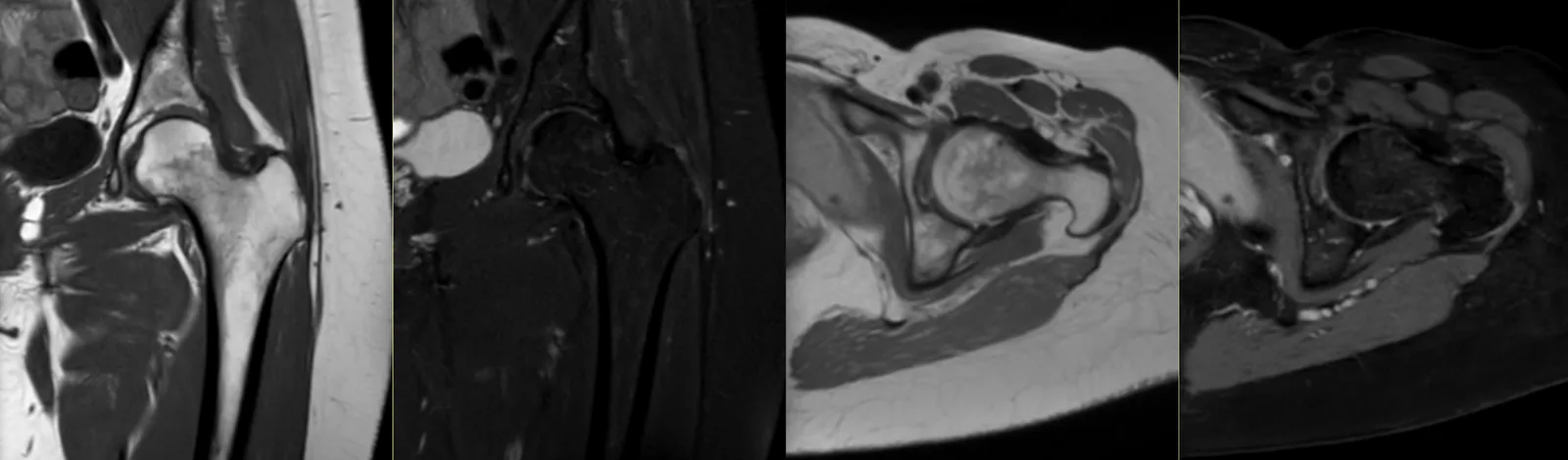
When it is requested
- Persistent hip or groin pain that a plain X-ray does not clarify.
- Suspected femoroacetabular impingement (CAM or Pincer).
- Suspected acetabular labral injury.
- Avascular necrosis (osteonecrosis) of the femoral head, including early stages.
- Suspected occult or stress fracture.
- Trochanteric bursitis or gluteal tendinopathy; athletic groin pain.
- Assessment before or after hip surgery.
What the exam is like
- 1Before
Reception and safety questionnaire
Arrive 15 minutes early and fill in the safety questionnaire, if you haven't already done so online.
- 2
Changing room
You undress to underwear with no metal parts; you are given a gown.
- 3
Positioning
You lie on your back, with your legs extended. One hip or the whole pelvis (both hips) may be studied, according to the request.
- 4
Inside the scanner
The hip sits at the centre of the magnet, so much of your body goes into the tunnel; your head is often near the opening.
- 520–30 min
Image acquisition
Several sequences with a characteristic knocking noise; you are given ear protection. In selected cases, an MR arthrogram (contrast in the joint) may be requested.
- 6
Finishing up
You get dressed and can leave without needing anyone to accompany you.
Preparation
- Fasting: not needed in general.
- Changing and clothing: as a rule, you undress to underwear with no metal parts — usually pants and socks. Depending on the area, the radiographer may allow you to keep some clothing on. You are given a gown.
- Devices and implants: note on the questionnaire any pacemaker, neurostimulator and hip replacement (does not prevent the exam, but produces artefacts near the implant).
- MR arthrogram: when indicated, it involves an injection of contrast into the joint by a doctor, before the exam.
- Claustrophobia: much of your body goes into the tunnel. If you are anxious, contact us before the day of the exam.
Results
The report is written by a radiologist from the images. The report is available as a PDF (by email or printed) and the images on CD and on CRMA's imaging platform, to take to your treating doctor — orthopaedic surgeon, physiatrist, family doctor or other.