
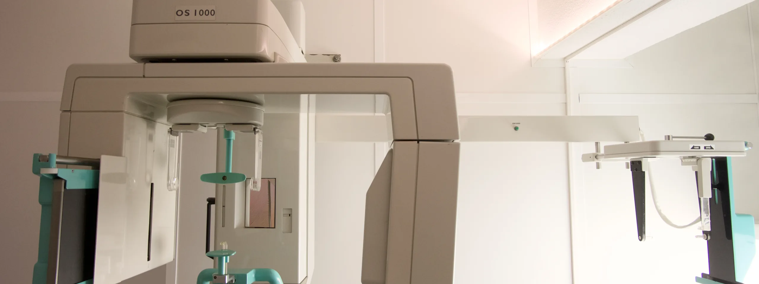
What does an orthopantomogram assess?
- Teeth — including impacted teeth and wisdom teeth.
- Mandible and maxilla — the supporting bone and the relationship between the dental arches.
- Temporomandibular joints and maxillary sinuses, in an overall view.
- Frontal and lateral cephalometric X-rays — calibrated views for cephalometric analysis, in orthodontics.
When the dentist needs a three-dimensional assessment — for example, for implants — CRMA also performs a CT Dental Scan, with 3D reconstructions.
When it is requested
- Overall assessment of the dentition — a first appointment or a dental treatment plan.
- Wisdom teeth — their position and relationship to neighbouring structures.
- Orthodontics — together with the cephalometric X-ray.
- Implant planning — often complemented by a CT Dental Scan.
- Assessment before oral surgery.
What the exam is like
- 1Before
Reception
Arrive with a referral from your dentist or treating doctor. No prior preparation.
- 2
Positioning
You stand at the equipment, with your chin resting on a support and biting gently on a disposable bite piece to hold the position.
- 3Seconds
Acquisition
The arm of the equipment rotates slowly around your head as it captures the image — you simply stay still for a few seconds.
- 4
Finishing up
You resume your day straight away, with no restrictions.
Preparation
- Preparation: none needed.
- Metal objects: remove earrings, facial piercings, necklaces, hair grips, glasses and removable dentures.
- Pregnancy or suspected pregnancy: always tell the team before the exam.
Results
The report is written by a radiologist. The report is available as a PDF (by email or printed) and the images on CRMA's imaging platform, to take to your treating doctor. With written authorisation, they can be sent directly to the referring doctor.