
What is assessed in a dental scan?
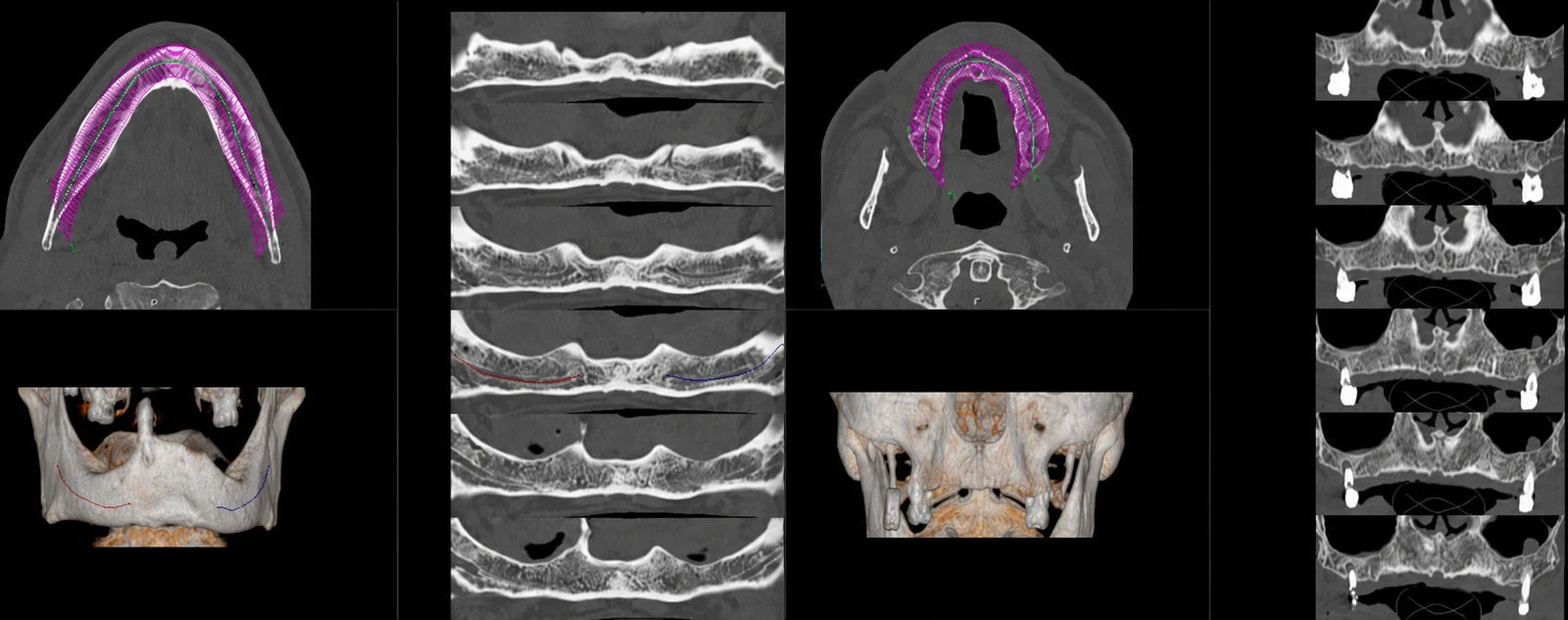
A dental scan allows detailed assessment of the jawbone — the maxilla (upper jaw) and the mandible (lower jaw) —, including the height, thickness and quality of the available bone, information that is essential for planning dental implants. It also allows study of impacted teeth (for example, wisdom teeth), their relationship with the inferior alveolar nerve canal and with the maxillary sinuses, and lesions of the jaws, such as cysts.
The images include 3D reconstructions and slices oriented along the dental arches, in the format that the dentist, the implantologist or the maxillofacial surgeon use to plan treatment. The report is produced by a radiologist; the clinical interpretation and the treatment decision rest with the referring doctor.
When it is requested
- Planning dental implants — assessment of the available bone and of the structures to preserve.
- Impacted teeth — the position of wisdom teeth or other unerupted teeth and their relationship with the alveolar nerve.
- Study of jaw lesions — cysts and other changes, according to clinical guidance.
- Pre-surgical assessment in oral and maxillofacial surgery.
- Relationship of the teeth with the maxillary sinuses — including sinus disease of dental origin.
- Assessment after bone grafts or other procedures, according to guidance.
What the exam is like
- 1Before
Reception
Arrive 15 minutes early, with your dentist's referral and any previous exams (e.g. an orthopantomogram), if you have them.
- 2
Preparation
You remove removable metal dentures and piercings from the face and mouth area, as well as earrings, hairpins and glasses.
- 3
Positioning
Lying on your back on the table, with your head in a dedicated support and your chin aligned. You may be asked not to swallow or move your mouth during acquisition.
- 45–10 min
Image acquisition
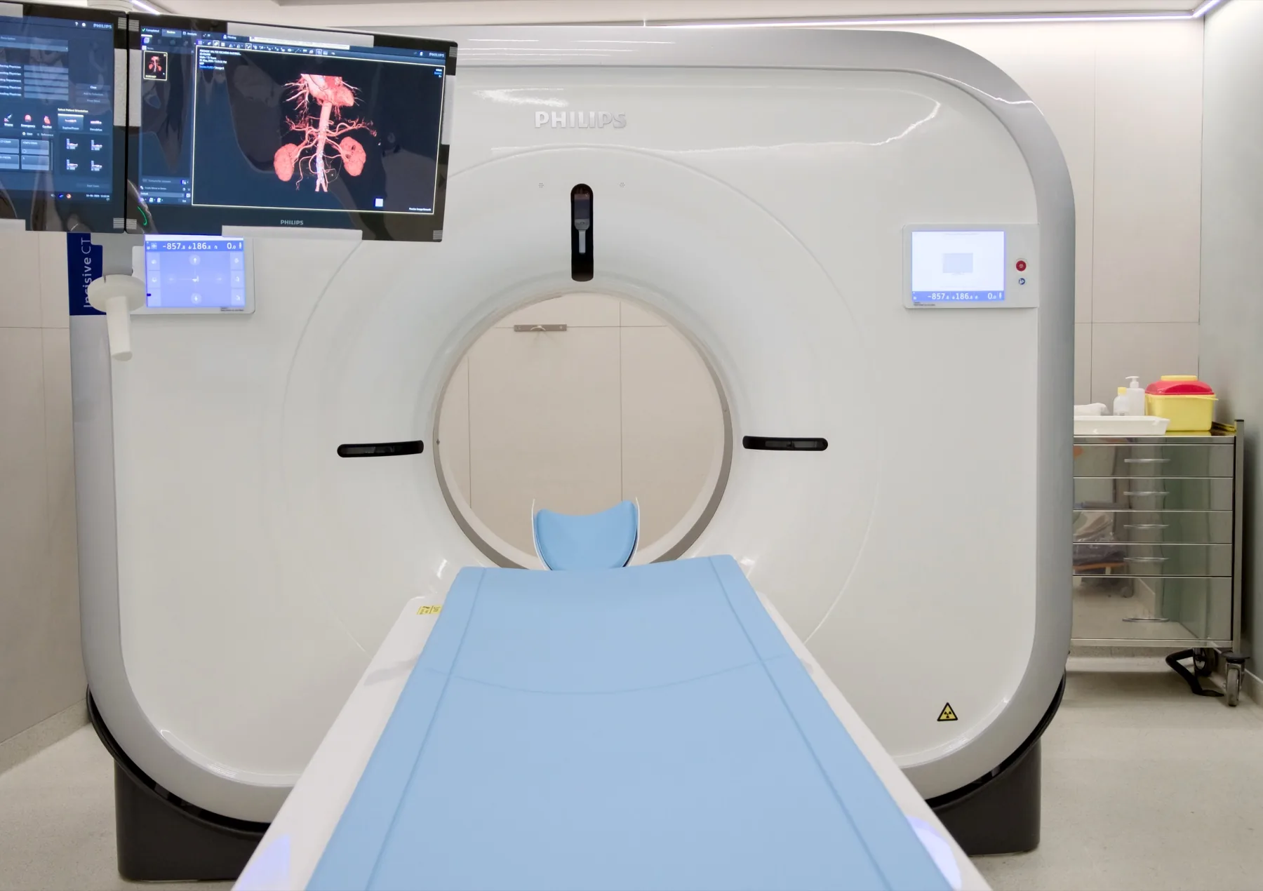
The table moves through the scanner's open ring; acquisition takes just seconds.
- 5
Finishing
You can leave, drive and resume your normal day straight away. The 3D reconstructions are prepared after the exam.
Preparation
- Fasting: not needed.
- Medication: keep your regular medication.
- Dentures and piercings: remove removable metal dentures and piercings from the face and mouth area at the time of the exam. Fixed dentures and existing implants do not prevent the exam — let us know about them when booking.
- Metal objects: remove earrings, hairpins and glasses.
- Clothing: as a rule, you keep your own clothes on.
- Pregnancy: CT uses X-rays. If you are or think you may be pregnant, always let us know before the exam.
- Documents: your dentist's/doctor's referral, insurer/scheme card (if applicable), previous exams (orthopantomogram, CT).
Results
The report is produced by a radiologist from the images. The report is available as a PDF (by email or printed) and the images — including the 3D reconstructions and the slices oriented along the dental arches — on CD and on CRMA's imaging platform, in the format your dentist uses for planning.