
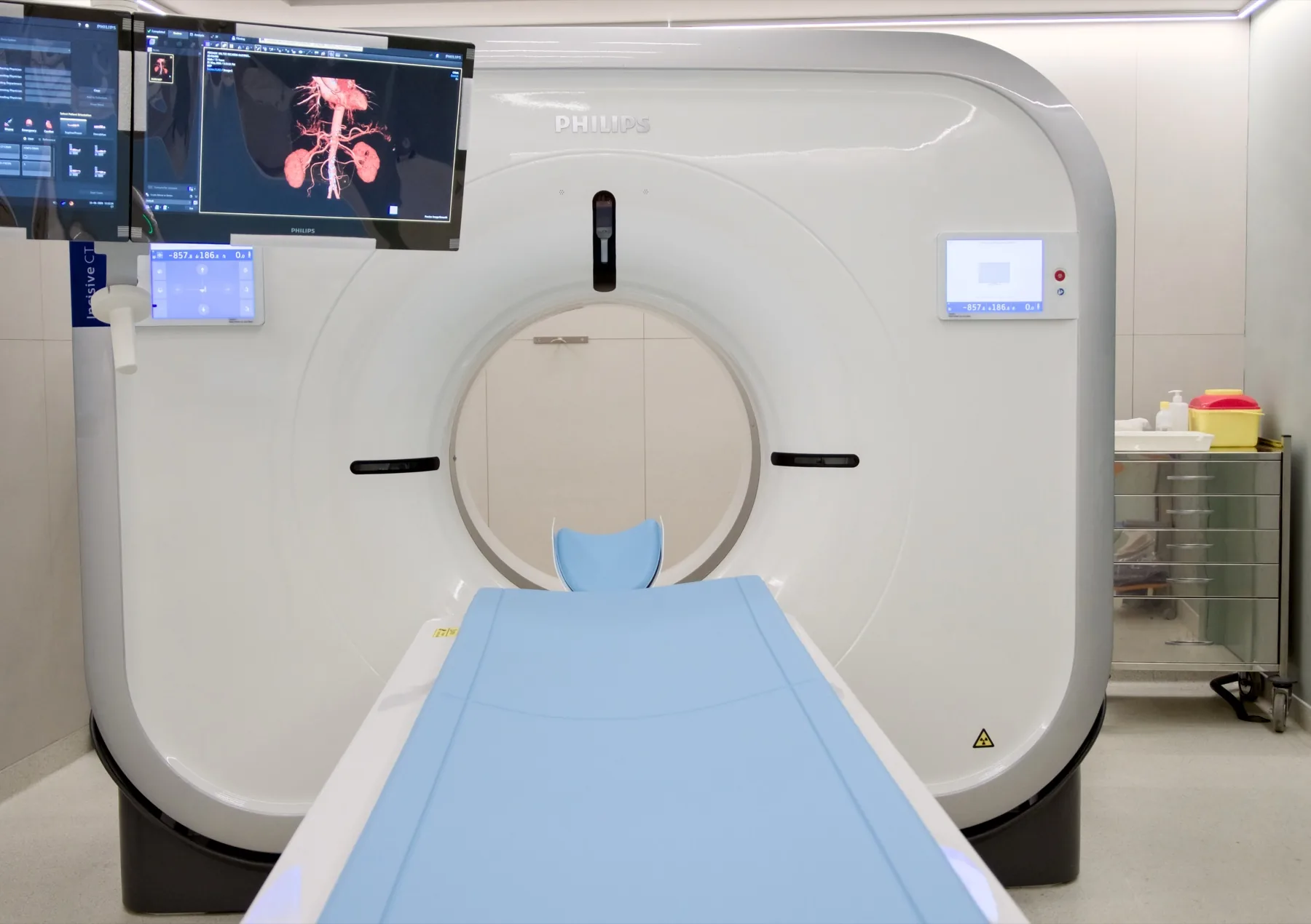
What is assessed in a lumbar spine CT scan?
A lumbar spine CT scan allows assessment of the lumbar vertebrae and the sacrum — the vertebral bodies, the posterior arches and the facet joints —, the spinal canal and the intervertebral discs, with a bone detail that is this exam's strong point. It also allows assessment of osteosynthesis hardware (screws, rods) after spinal surgery.
CT and magnetic resonance imaging are complementary exams: for the study of the discs, the nerves and the spinal cord, MRI is usually the exam of choice; CT stands out for its bone detail and is the alternative when MRI is not possible. The choice always rests with the referring doctor. The report is produced by a radiologist; the clinical interpretation rests with your doctor.
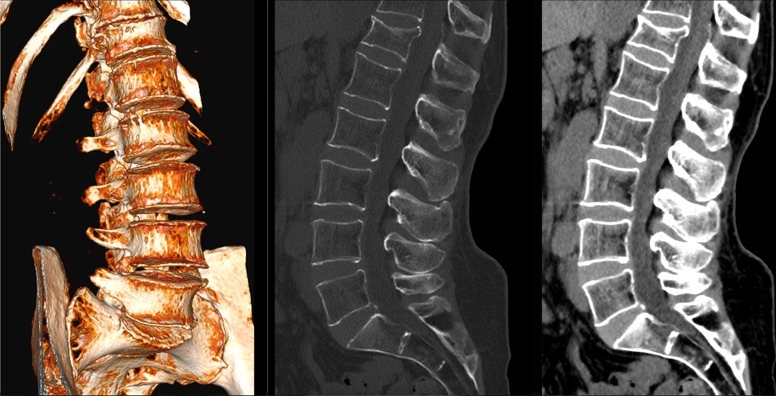
When it is requested
- Assessment of vertebral fractures — traumatic or fragility fractures (osteoporosis).
- Low back pain under investigation, when MRI is not possible or as a complement.
- Degenerative changes — facet joint osteoarthritis, spinal canal narrowing of bony origin.
- Spondylolysis and spondylolisthesis — vertebral defects and slippage.
- Assessment after spinal surgery — positioning and integrity of the osteosynthesis hardware.
- Planning of surgery or of image-guided procedures.
- When MRI is contraindicated — some pacemakers and devices, severe claustrophobia.
What the exam is like
- 1Before
Reception
Arrive 15 minutes early, with your referral and any previous spine exams, if you have them.
- 2
Preparation
You remove only the metal objects from the lumbar area: belt, fasteners, piercings. As a rule, you keep your own clothes on.
- 3
Positioning
Lying on your back on the table, with your legs slightly raised on a support, for comfort of the lumbar region.
- 45–10 min
Image acquisition
The table moves through the scanner's open ring. You just need to keep still for a few brief moments.
- 5
Finishing
You can leave, drive and resume your normal day straight away.
Preparation
- Fasting: not needed.
- Contrast: rarely needed. Even when it is used, fasting is not required; only the kidney-function recommendations apply (recent blood tests from the last 3 to 6 months, if you have them) — we'll let you know when booking.
- Medication: keep your regular medication.
- Metal objects: remove only those from the area being studied — belt, fasteners, piercings.
- Clothing: as a rule, you keep your own clothes on, provided they have no metal parts in the lumbar area.
- Surgical hardware: screws, rods or prostheses in the spine do not prevent the CT scan — mention them when booking, they help us plan the exam.
- Pregnancy: CT uses X-rays. If you are or think you may be pregnant, always let us know before the exam.
- Pain when lying down: if you find it hard to lie on your back, tell us — the positioning is adjusted with you.
- Documents: referral, insurer/scheme card (if applicable), previous spine exams (X-ray/CT/MRI).
Results
The report is produced by a radiologist from the images. The report is available as a PDF (by email or printed) and the images on CD and on CRMA's imaging platform, to take to your doctor.