
What is a CT angiography (CTA)?
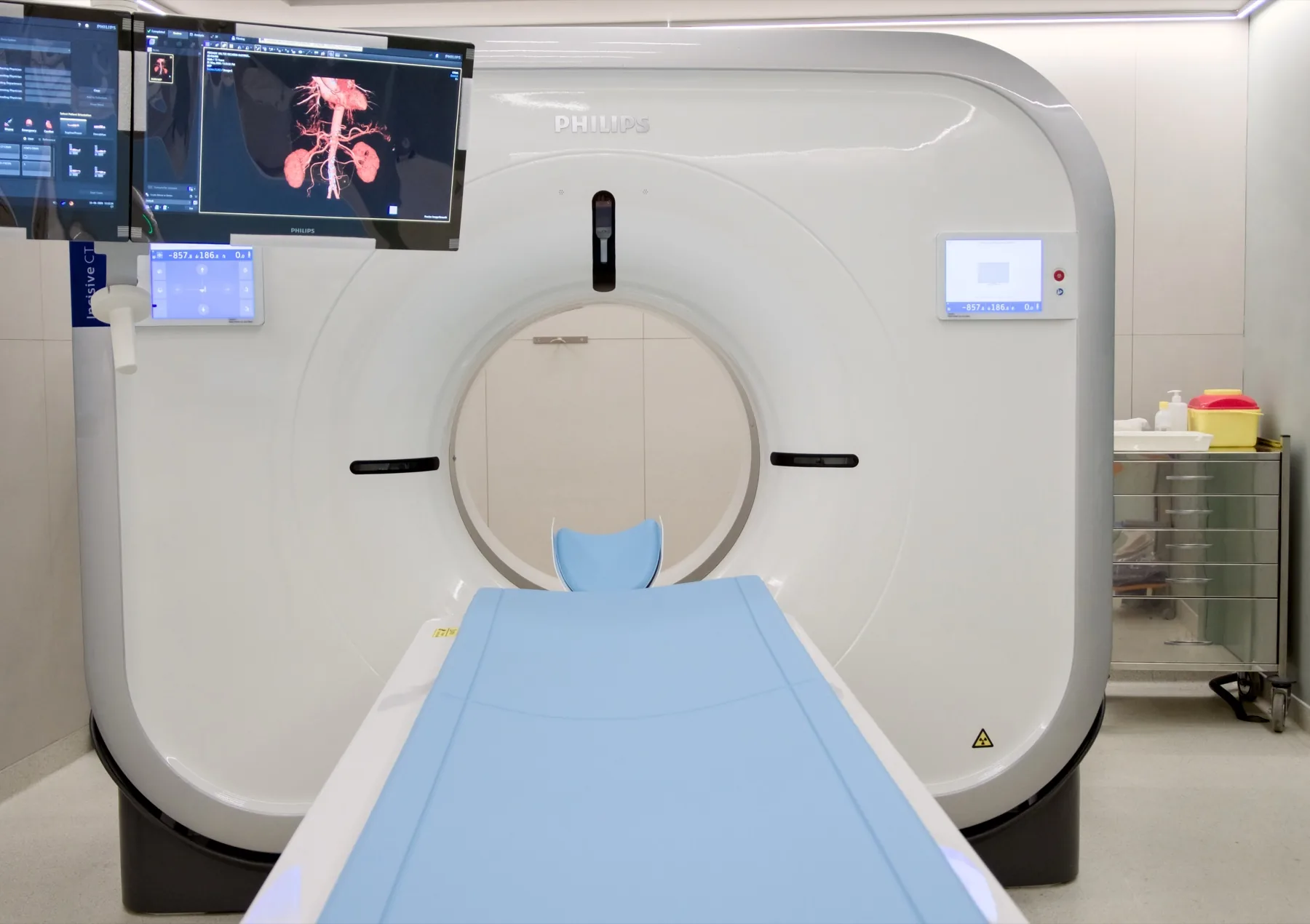
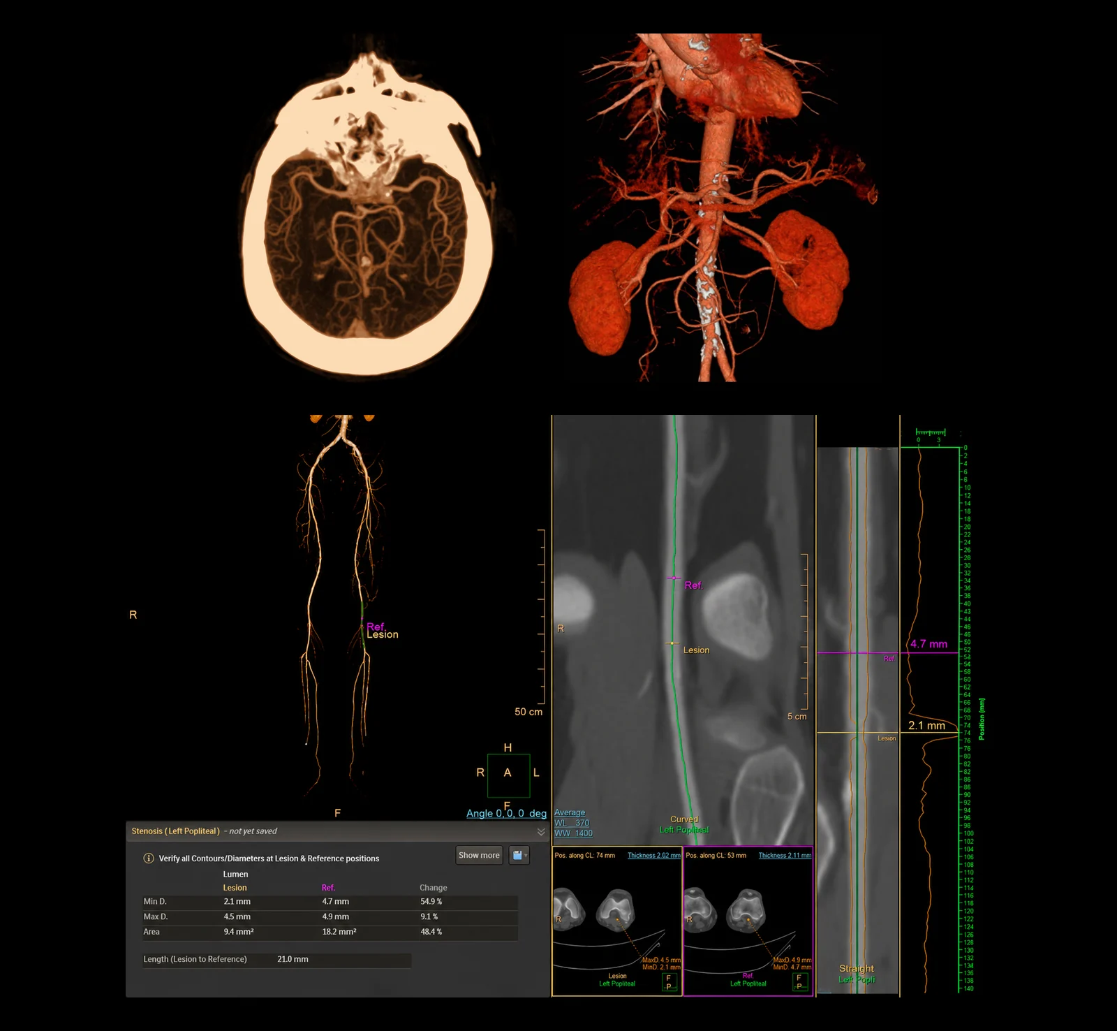
A CT angiography (CTA — also known as angio-CT) is a study aimed specifically at the blood vessels. It differs from a conventional CT scan by: synchronisation with the contrast (bolus tracking, which triggers acquisition at the ideal density); dedicated post-processing (MIP, MPR and VR reconstructions); optimised low-voltage protocols.
The Philips Incisive 128-slice scanner allows acquisitions with a high pitch and an optimised dose.
When it is requested
- Acute suspicion: pulmonary thromboembolism (CTPA), aortic dissection, ruptured aneurysm.
- Chronic vascular disease: atherosclerotic stenoses (carotid, renal, peripheral), stable aneurysms, follow-up after stenting.
- Pre-surgical mapping: before elective vascular surgery (endarterectomy, bypass, EVAR/TEVAR), before transplantation.
Territories available at CRMA
- CTPA — Pulmonary: first-line for pulmonary thromboembolism (ACR Appropriateness 2022).
- Thoracic and abdominal aorta: dissection, aneurysm, follow-up after EVAR/TEVAR.
- Carotid and vertebral arteries: carotid stenosis, dissection, anomalies of the supra-aortic arches.
- Head (intracranial vessels): aneurysm, AVM, follow-up after endovascular treatment.
- Renal arteries: refractory hypertension, fibromuscular dysplasia, pre-transplant mapping.
- Iliac arteries: occlusive disease, pre-TAVI assessment, pelvic mapping.
- Lower limbs (runoff): peripheral arterial disease, critical ischaemia, before bypass.
- Upper limbs: thoracic outlet syndrome, subclavian thrombosis, pre-fistula mapping.
What the exam is like
The exam is fast — generally between 5 and 15 minutes from entering the room. The image acquisition itself takes seconds to a few minutes.
- 1Before
Reception
Arrive 15 minutes early. We confirm the booking, the referral and the funding scheme, and review the relevant clinical history (allergies, kidney function, medication, pregnancy).
- 2
Cannula for the contrast
A cannula is placed in your arm (usually at the crook of the elbow); it stays in place during the exam and is removed at the end.
- 3
Positioning
Lying on your back on the scanner table, after removing metal objects from the area being studied. The radiographer follows the whole exam from the control room, in contact with you.
- 4
Contrast injection
The iodinated contrast is injected by an automatic pump at a controlled rate, synchronised with acquisition through bolus tracking. You may feel a diffuse warmth through the body during the injection — this is normal and passing.
- 5Seconds
Image acquisition
You stay still for a few seconds; for studies of the chest and abdomen, you are asked to hold your breath briefly.
- 6
Finishing
The cannula is removed. You can leave, drive and resume your normal day — we recommend drinking water over the following hours.
Preparation
- Fasting: not required. Have a light meal before you come and drink water freely up to the time of your exam.
- Hydration: drink water in the hours before and after.
- Kidney function: no blood test is required. If you have recent blood tests (last 3 to 6 months) with creatinine/kidney function, it's advisable to bring them. If you have known kidney disease, let us know when booking — the use of contrast is assessed case by case.
- Allergies to iodine / previous contrast: let us know when booking; pre-medication is possible.
- Medication: keep your regular medication — including your diabetes and blood pressure medication, and metformin — unless your doctor advises otherwise.
- Breastfeeding: always let us know. Breastfeeding may continue as normal after the contrast is given (ACR/ESUR recommendation).
Results
The report is produced by a radiologist from the images. The report is available as a PDF (by email or printed) and the images on CD and on CRMA's imaging platform, to take to your treating doctor.