
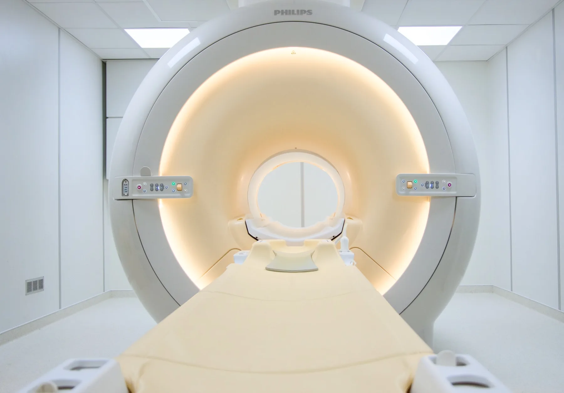
What is assessed in a knee MRI?
Knee magnetic resonance imaging (MRI) allows assessment of all the structures that make up the joint — the menisci, cruciate ligaments (anterior and posterior), collateral ligaments, articular cartilage, tendons (patellar, quadriceps, pes anserinus), bursae, bones (femur, tibia, fibula, patella) and the soft tissue around the joint.
Because it uses a magnetic field rather than X-rays, MRI involves no ionising radiation and offers resolution that is particularly useful for soft-tissue injuries.
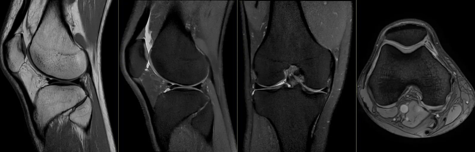
When it is requested
- Persistent knee pain that a plain X-ray does not clarify.
- Suspected meniscal injury after a twist, fall or sport.
- Suspected ligament injury (anterior cruciate, posterior cruciate, collateral ligaments), particularly after a sprain with a feeling of instability.
- Cartilage injury or chondropathy.
- Assessment before surgery (arthroscopy, cruciate ligament reconstruction, joint replacement).
- Follow-up after surgery.
- Suspected occult bone injury that does not show on an X-ray.
What the exam is like
- 1Before
Reception and safety questionnaire
Arrive 15 minutes early and fill in the safety questionnaire, if you haven't already done so online.
- 2
Changing room
Remove any metal objects; if your clothing from the waist down has no metal parts, you may keep it on. You may be given a gown.
- 3
Positioning
You lie on your back, with a flexible surface coil wrapped around the knee; support cushions help minimise movement.
- 4
Inside the scanner
Most of your body stays outside the tunnel — your head is often outside or near the opening. You stay in contact with the radiographer and are given an emergency call button.
- 520–30 min
Image acquisition
Several sequences with a characteristic knocking noise; you are given ear protection. Contrast is rarely needed; in selected cases, the doctor may request an MR arthrogram.
- 6
Finishing up
You get dressed and can leave without needing anyone to accompany you.
Preparation
- Fasting: not needed, even when contrast is used — which is rare for a knee MRI.
- Clothing: comfortable, with no metal parts. Loose shorts are welcome.
- Osteosynthesis hardware: most modern hardware is MRI-compatible. If you have had surgery, bring the documentation for the implanted hardware.
- Knee replacement: does not prevent the exam. It may produce artefacts in the area of the implant.
- Documents: your referral, your insurer or subsystem card, and any previous knee exams.
Results
The report is written by a radiologist from the images. The report is available as a PDF (by email or printed) and the images on CD and on CRMA's imaging platform, to take to your treating doctor — orthopaedic surgeon, physiatrist, family doctor or other.