What is a CT scan?
Computed tomography (CT — also known as a CAT scan) is a medical imaging technique that uses X-rays combined with computer processing to produce cross-sectional images of the body.
It allows precise assessment of the bony structures, blood vessels, internal organs and acute injuries. It is often requested in urgent outpatient settings, trauma, oncology, neurology and abdominal conditions.
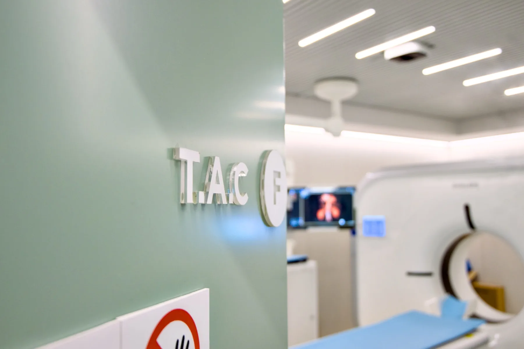
CRMA has a Philips Incisive CT 128-slice scanner (installed in 2026), with a complete cardiac package and optimised low-dose protocols.
How it's done
The exam is fast — in general between 5 and 20 minutes. Image acquisition itself takes from seconds to a few minutes. For exams with contrast, a cannula is placed in the arm; you may feel a passing sensation of warmth — this is normal.
Preparation
- Fasting: not required, even with contrast. Have a light meal before you come and drink water freely up to the time of your exam.
- Kidney function: we don't require blood tests; if you have recent tests (last 3 to 6 months) with creatinine/kidney function, it's advisable to bring them. If you have known kidney disease, let us know when booking.
- Medication: keep your regular medication — including your diabetes and blood pressure medication, and metformin — unless your doctor advises otherwise.
- Allergies: let us know when booking about any previous reactions to iodinated contrast — each case is assessed individually by the radiologist.
- Oral contrast: for some chest, abdominal or pelvic studies, according to the clinical information — it is provided at CRMA; in those cases, arrive at least 30 minutes before your appointment time.
- Hydration: we recommend drinking water throughout the day after an exam with contrast.
Results
The report is produced by a radiologist from the images. The report is available as a PDF (by email or printed) and the images on CD and on CRMA's imaging platform, to take to your doctor.
Variants of CT
Eleven CT exams have their own page, with preparation, step-by-step details and frequently asked questions:
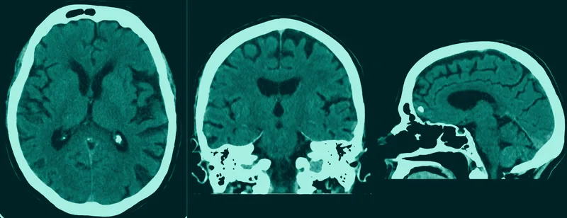
Head CT
Skull and brain — injury, acute settings.
Learn more →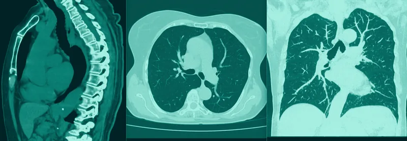
Chest CT
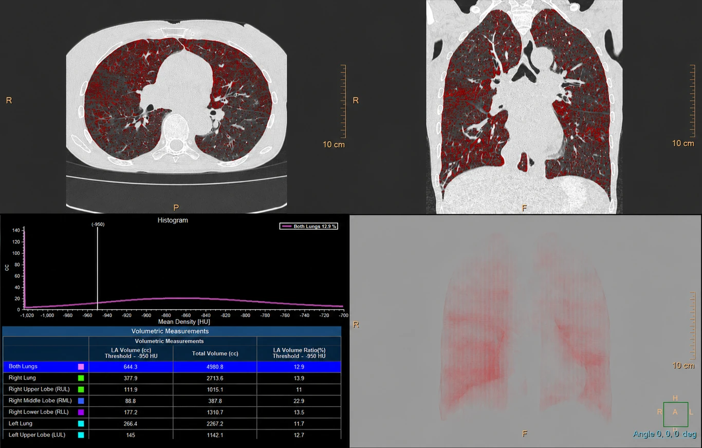
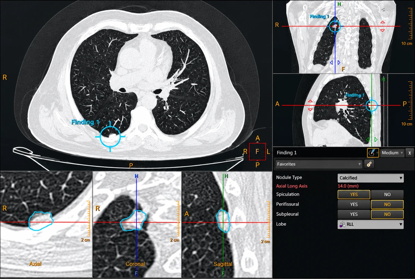
Lungs, mediastinum and pleura; quantification of emphysema and nodules.
Learn more →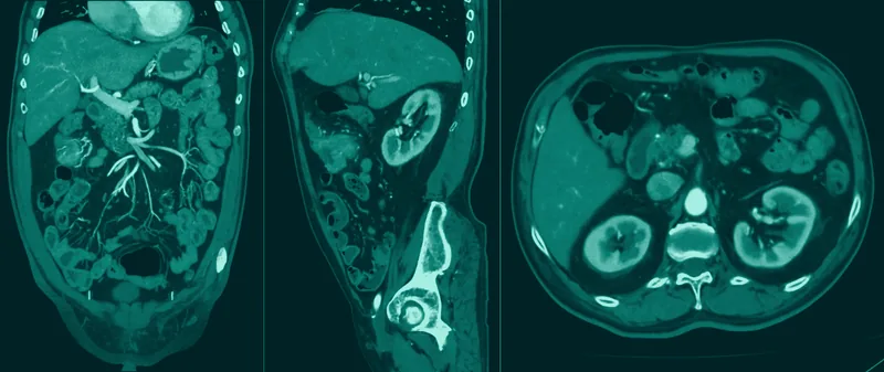
Abdominal and Pelvic CT
Liver, pancreas, kidneys, bowel and pelvic organs.
Learn more →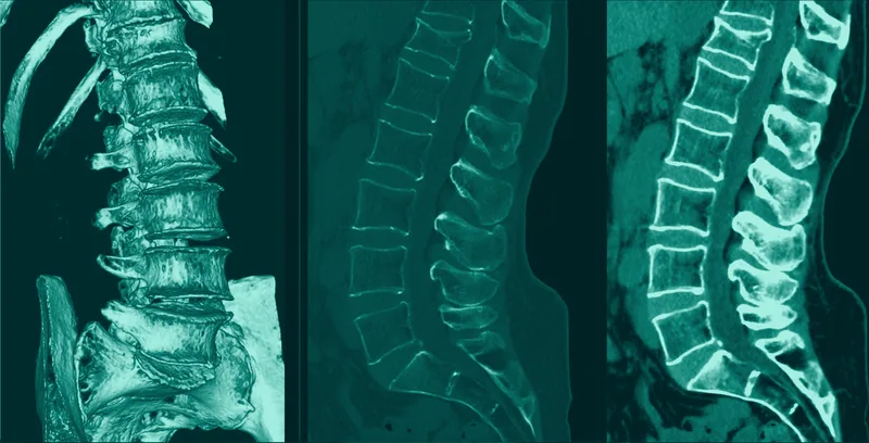
Lumbar Spine CT
Bone detail, fractures and surgical hardware.
Learn more →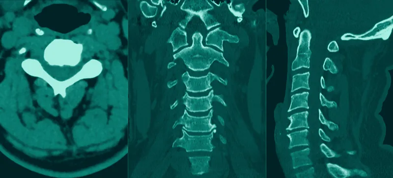
Cervical Spine CT
Vertebrae of the neck — injury and post-surgery.
Learn more →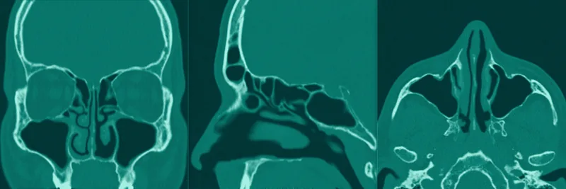
Paranasal Sinuses CT
Chronic sinusitis and nasosinus surgery.
Learn more →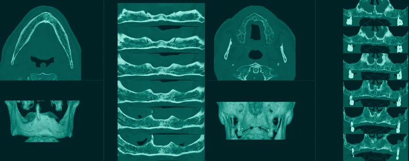
Dental Scan
Jaws in 3D — implant planning.
Learn more →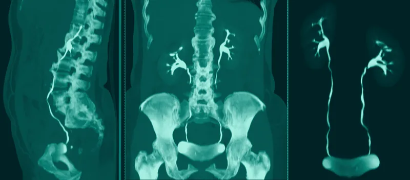
Uro-CT
Kidneys, ureters and bladder — stones and haematuria.
Learn more →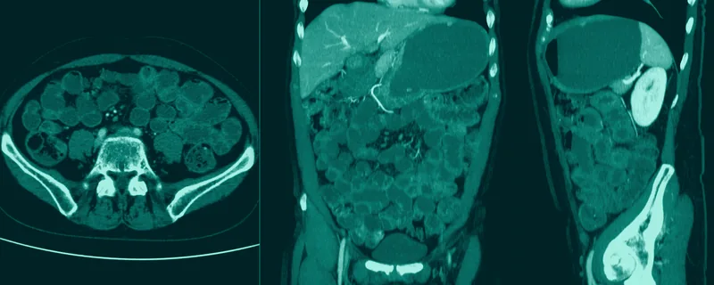
CT enterography
Small bowel — Crohn's disease.
Learn more →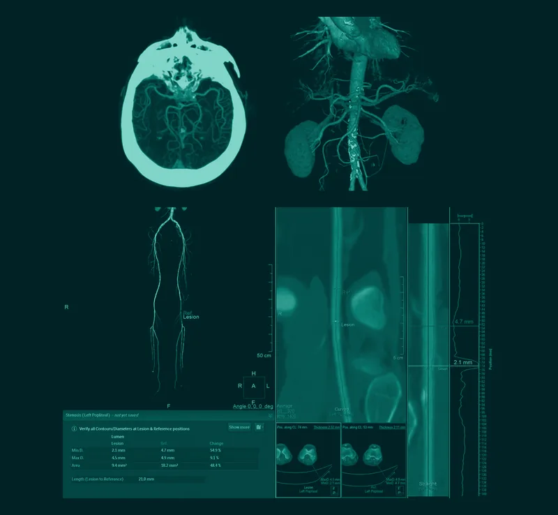
CT angiography
Vascular studies in 9 territories.
Learn more →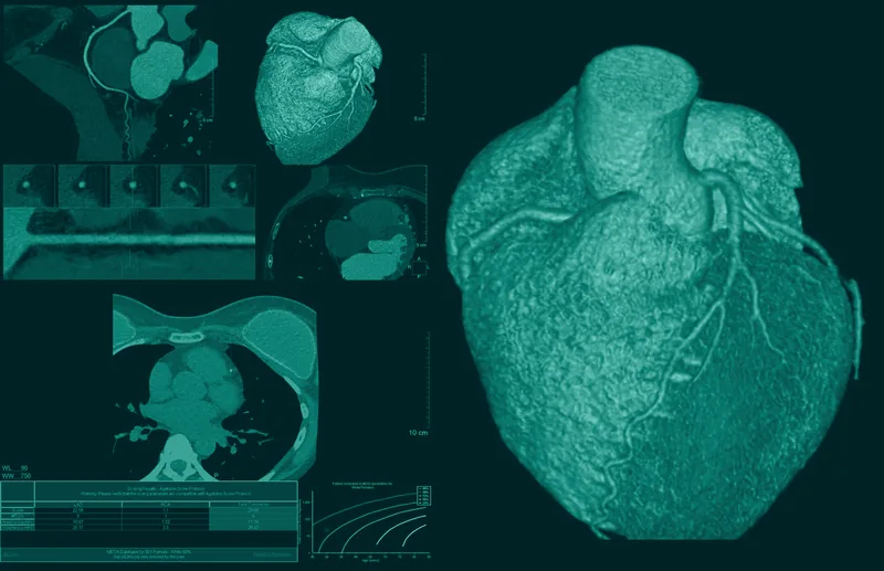
Cardiac CT angiography + CACS
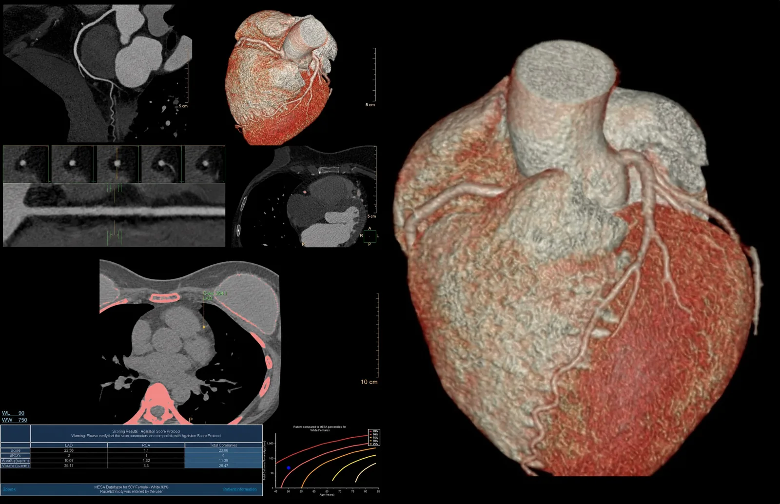
Coronary arteries and Calcium Score. New service 2026.
Learn more →Full list of variants by region — each exam is performed with a dedicated protocol, adjusted by the radiologist to the clinical indication:
- Head and neck: head (brain), pituitary gland, internal auditory canals, ears and mastoids, orbits, face, paranasal sinuses and jaws, nasopharynx, temporomandibular joints, jaws, tongue, larynx, neck, thyroid.
- Spine: cervical, thoracic, lumbar and lumbosacral, sacral, coccygeal, sacroiliac joints.
- Chest, abdomen and pelvis: chest, mediastinum, abdomen and pelvis, renal and/or adrenal, rectum, combined studies (chest + abdomen + pelvis), Uro-CT, CT enterography.
- Musculoskeletal: pelvis/hip joints, shoulder, elbow, knee(s), ankle, foot, lower limbs, CT arthrography.
- Dental Scan: both arches, maxilla or mandible, with 3D reconstructions.
- Vascular studies: CT angiography in 9 territories and Cardiac CT angiography + Calcium Score (CACS).