
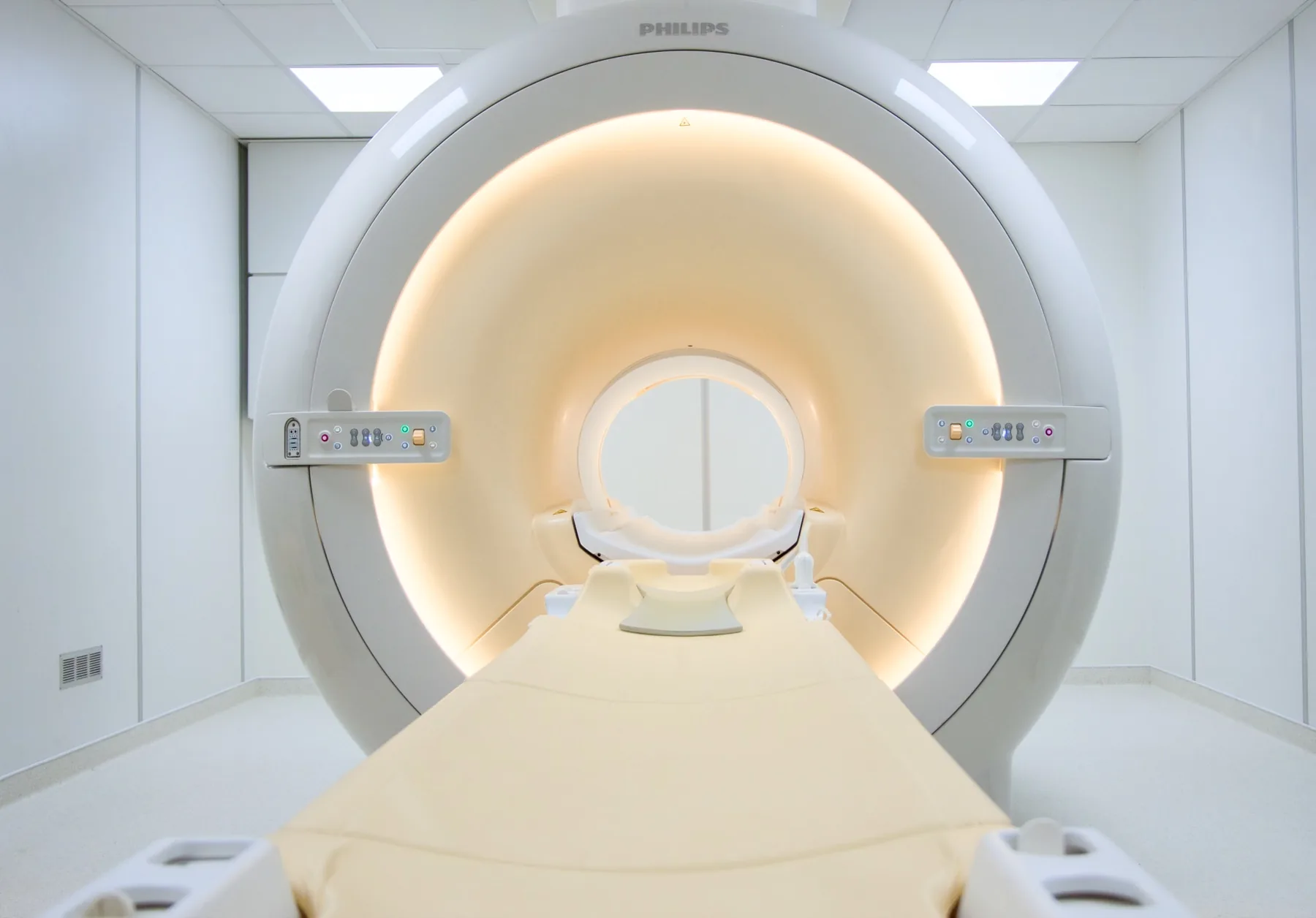
What is assessed in a lumbar spine MRI?
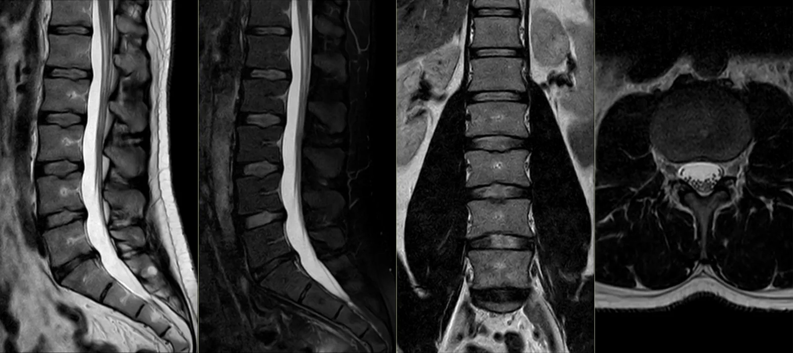
Lumbar spine magnetic resonance imaging (MRI) allows assessment of the structures of the lumbar and lumbosacral region — the vertebrae (L1 to L5 and the sacrum), the intervertebral discs, the spinal canal, the conus medullaris and the cauda equina, the nerve roots, the facet joints, the ligaments and the soft tissue around the spine.
Because it uses a magnetic field rather than X-rays, MRI involves no ionising radiation and offers resolution that is particularly useful for the soft tissues — discs, spinal cord and nerves — which often cannot be seen on a plain X-ray. The report is written by a radiologist; the clinical interpretation and the decision on the next step rest with your treating doctor.
When it is requested
- Persistent low back pain that a plain X-ray does not clarify.
- Sciatica (lumbar radicular pain) — pain that radiates to the buttock and leg, sometimes with tingling or loss of strength.
- Suspected disc herniation — to assess the discs and their relationship with the nerve roots.
- Suspected spinal canal stenosis — narrowing of the canal through which the nerves pass.
- Spondylolisthesis — slippage of one vertebra over another.
- Assessment before spinal surgery and follow-up after surgery.
- Inflammatory changes of the spine (for example, in the context of spondyloarthritis).
What the exam is like
- 1Before
Reception and safety questionnaire
Arrive 15 minutes before your appointment and fill in the safety questionnaire, if you haven't already done so online.
- 2
Changing room
You undress and stay in underwear with no metal parts; you are given a gown.
- 3
Positioning
You lie on your back (supine), usually with your legs supported on a cushion for greater comfort.
- 4
Inside the scanner
The lower back sits in the centre of the magnet, so much of your body — including your head — stays inside the tunnel. You stay in contact with the radiographer via a microphone and have an emergency call button.
- 520–30 min
Image acquisition
Several sequences, each with a characteristic noise; you are given ear protection. It is important to stay still during each sequence.
- 6
Finishing up
You get dressed and can leave without needing anyone to accompany you. Contrast (gadolinium) is rarely needed.
Preparation
- Fasting: not needed in general, even when contrast is anticipated.
- Changing and clothing: as a rule, you undress and stay in underwear with no metal parts — usually briefs and socks. Depending on the area, the radiographer may allow you to keep some clothing on. You are given a gown.
- Devices and implants: note on the safety questionnaire any pacemaker, neurostimulator, insulin pump, brain clips or osteosynthesis hardware in the spine.
- Documents: your referral, your insurer or subsystem card (if you have an agreement) and any previous spine exams.
- Claustrophobia: in a lumbar spine MRI your head stays inside the tunnel. If you are anxious, contact us before the day of the exam.
Results
The report is written by a radiologist from the images obtained. We let you know when it is ready, as agreed at booking. The report is available as a PDF (by email or printed) and the images on CD and on CRMA's imaging platform, to take to your treating doctor — orthopaedic surgeon, neurosurgeon, physiatrist, family doctor or other.