
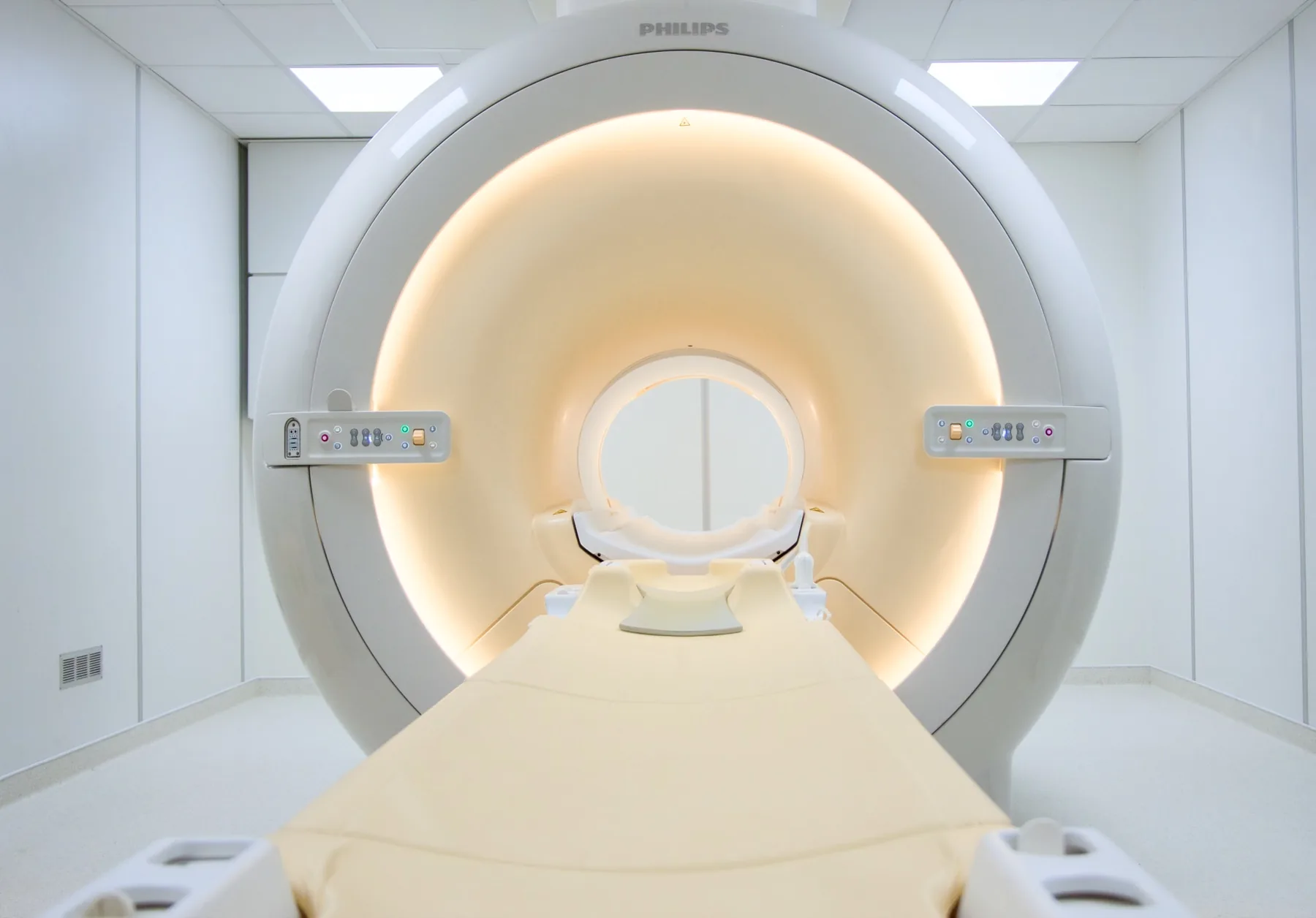
What is assessed in a cervical spine MRI?
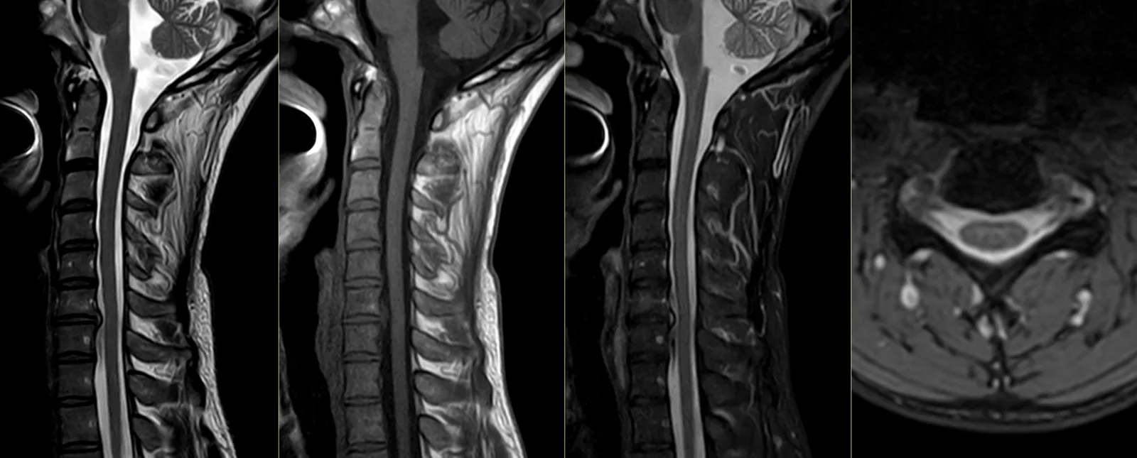
Cervical spine MRI allows assessment of the structures of the neck — the cervical vertebrae (C1 to C7), the intervertebral discs, the cervical spinal cord, the nerve roots, the spinal canal and the cervical joints.
Unlike the lumbar spine — where the spinal cord ends higher up — in the cervical spine the spinal cord runs through the whole region. This makes cervical spine MRI particularly useful for assessing the relationship between the vertebrae/discs and the spinal cord itself. Because it uses a magnetic field, it involves no ionising radiation.
When it is requested
- Persistent neck pain that a plain X-ray does not clarify.
- Cervicobrachial pain — pain that radiates from the neck into the shoulder, arm or hand, sometimes with tingling or loss of strength.
- Suspected cervical disc herniation.
- Suspected cervical myelopathy — signs of spinal cord involvement, such as changes in gait or hand dexterity.
- Cervical canal stenosis.
- Assessment before or after cervical spine surgery.
- Study of the spinal cord in demyelinating diseases (e.g. multiple sclerosis), often together with a brain MRI.
What the exam is like
- 1Before
Reception and safety questionnaire
Arrive 15 minutes early and fill in the safety questionnaire, if you haven't already done so online.
- 2
Changing room
You remove your clothing and stay in underwear with no metal parts; take off necklaces, earrings and hair grips. You are given a gown.
- 3
Positioning
You lie on your back, with your head and neck resting on a dedicated coil.
- 4
Inside the scanner
Your head and neck are inside the tunnel. You should keep your neck still and avoid swallowing repeatedly during each sequence.
- 520–30 min
Image acquisition
Several sequences, each with a characteristic noise; you are given ear protection and stay in contact with the radiographer.
- 6
Finishing up
You get dressed and can leave without needing anyone to accompany you. Contrast is uncommon.
Preparation
- Fasting: not needed in general, even when contrast is anticipated.
- Changing room and clothing: as a rule, you remove your clothing and stay in underwear with no metal parts — usually underwear and socks. Depending on the area, the radiographer may allow you to keep some clothing on. You are given a gown.
- Devices and implants: note them on the safety questionnaire (pacemaker, neurostimulator, clips, cochlear implant, cervical hardware).
- Documents: your referral, your insurer or subsystem card (if you have an agreement), and any previous cervical spine exams.
- Claustrophobia: your head and neck are inside the tunnel. If you are anxious about this, contact us before the day of the exam.
Results
The report is written by a radiologist from the images. The report is available as a PDF (by email or printed) and the images on CD and on CRMA's imaging platform, to take to your treating doctor — orthopaedic surgeon, neurosurgeon, neurologist, physiatrist or other.