
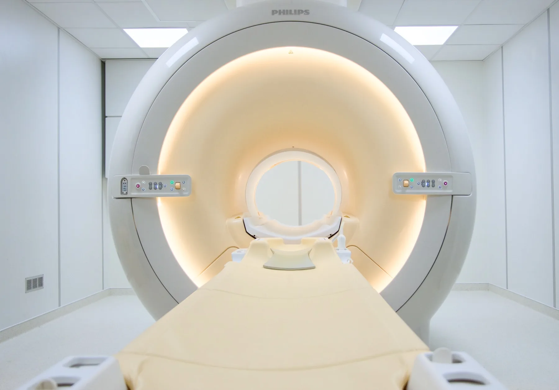
What is assessed in a shoulder MRI?
A shoulder MRI allows assessment of the structures of the joint — the rotator cuff tendons (supraspinatus, infraspinatus, subscapularis and teres minor), the long head of the biceps tendon, the labrum, the cartilage, the capsule, the subacromial-subdeltoid bursa and the bones (humerus, scapula and acromioclavicular joint).
Because it uses a magnetic field rather than X-rays, MRI involves no ionising radiation and offers resolution that is particularly useful for the soft tissues — tendons, labrum and cartilage. The report is written by a radiologist; the clinical interpretation rests with your treating doctor.
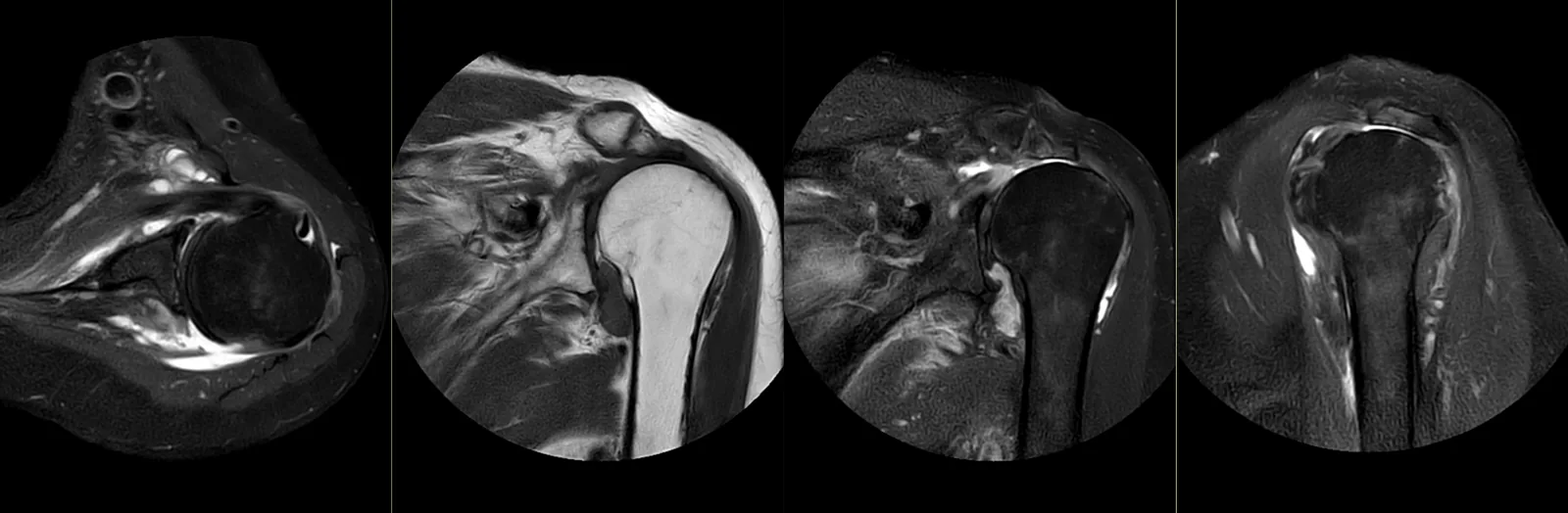
When it is requested
- Chronic shoulder pain that a plain X-ray does not clarify.
- Suspected rotator cuff injury (tear), particularly with loss of strength.
- Subacromial impingement (impingement syndrome), tendinitis and cuff tendinopathy.
- Suspected labral injury (e.g. SLAP) and instability or recurrent dislocation.
- Adhesive capsulitis ("frozen shoulder").
- Assessment before or after shoulder surgery.
What the exam is like
- 1Before
Reception and safety questionnaire
Arrive 15 minutes early and fill in the safety questionnaire, if you haven't already done so online.
- 2
Changing room
You undress and stay in underwear with no metal parts; you are given a gown.
- 3
Positioning
You lie on your back, with the arm being examined by your side and a flexible surface coil over the shoulder.
- 4
Inside the scanner
As the shoulder sits at the centre of the magnet, part of your head and trunk enter the tunnel. You stay in contact with the radiographer.
- 520–30 min
Image acquisition
Several sequences with a characteristic noise; you are given ear protection. In selected cases, the doctor may request an MR arthrogram (contrast into the joint).
- 6
Finishing up
You get dressed and can leave without needing anyone to accompany you.
Preparation
- Fasting: not needed, even with contrast. Only for exams with sedation (rare) do we ask for 6 hours without solid food, with clear fluids allowed until 2 hours before.
- Changing room and clothing: as a rule, you undress and stay in underwear with no metal parts — usually underwear and socks. Depending on the area, the radiographer may allow you to keep some clothing on. You are given a gown.
- Devices and implants: note any pacemaker, neurostimulator or osteosynthesis hardware in the shoulder on the questionnaire.
- MR arthrogram: when indicated, it involves an injection of contrast into the joint by a doctor, before the exam.
- Claustrophobia: part of your head and trunk enter the tunnel. If you feel anxious, contact us before the day of the exam.
Results
The report is written by a radiologist from the images. The report is available as a PDF (by email or printed) and the images on CD and on CRMA's imaging platform, to take to your treating doctor — orthopaedic surgeon, physiatrist, family doctor or other.