
What is assessed in a head CT scan?
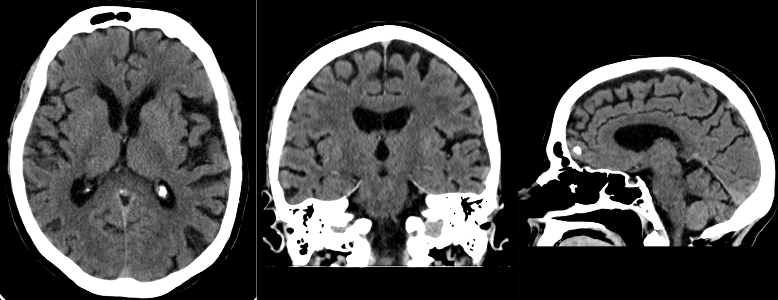
A head CT scan allows assessment of the skull bones and the brain — the cerebrum, the cerebellum and the brainstem —, as well as the ventricles and the cerebrospinal fluid spaces. It is particularly useful in the assessment of fractures and of acute intracranial haemorrhage, where speed of execution makes a difference. When administered, iodinated contrast allows certain lesions to be studied in more detail, according to the clinical indication; combined with CT angiography, it also studies the vessels of the head and neck.
CT and magnetic resonance imaging are complementary exams: CT stands out for its speed and its bone detail; MRI, for the detail of the nervous-system tissues. The choice always rests with the referring doctor. The report is produced by a radiologist; the clinical interpretation rests with your doctor.
When it is requested
- After head injury — assessment of fractures and haemorrhage.
- Sudden, severe headache, or one with features different from usual.
- Initial assessment of suspected stroke — above all to assess for the presence of haemorrhage.
- Acute neurological changes — confusion, loss of strength, speech changes.
- Dizziness, vertigo or balance problems, according to clinical guidance.
- Follow-up of known conditions — hydrocephalus, shunt valves, post-operative monitoring.
- Targeted study of the bony structures of the skull.
- When MRI is not possible — incompatible devices, severe claustrophobia or another contraindication.
What the exam is like
- 1Before
Reception
Arrive 15 minutes early. You do not need to fast, even if the exam is with contrast; if you have recent blood tests with kidney function (last 3 to 6 months), it's advisable to bring them.
- 2
Preparation
You remove only the metal objects from the head and neck area: hairpins, earrings, piercings, glasses, removable dentures, hearing aids. As a rule, you keep your own clothes on.
- 3
Positioning
Lying on your back on the table, with your head in a dedicated support. The radiographer follows the whole exam from the control room, in contact with you.
- 45–10 min
Image acquisition
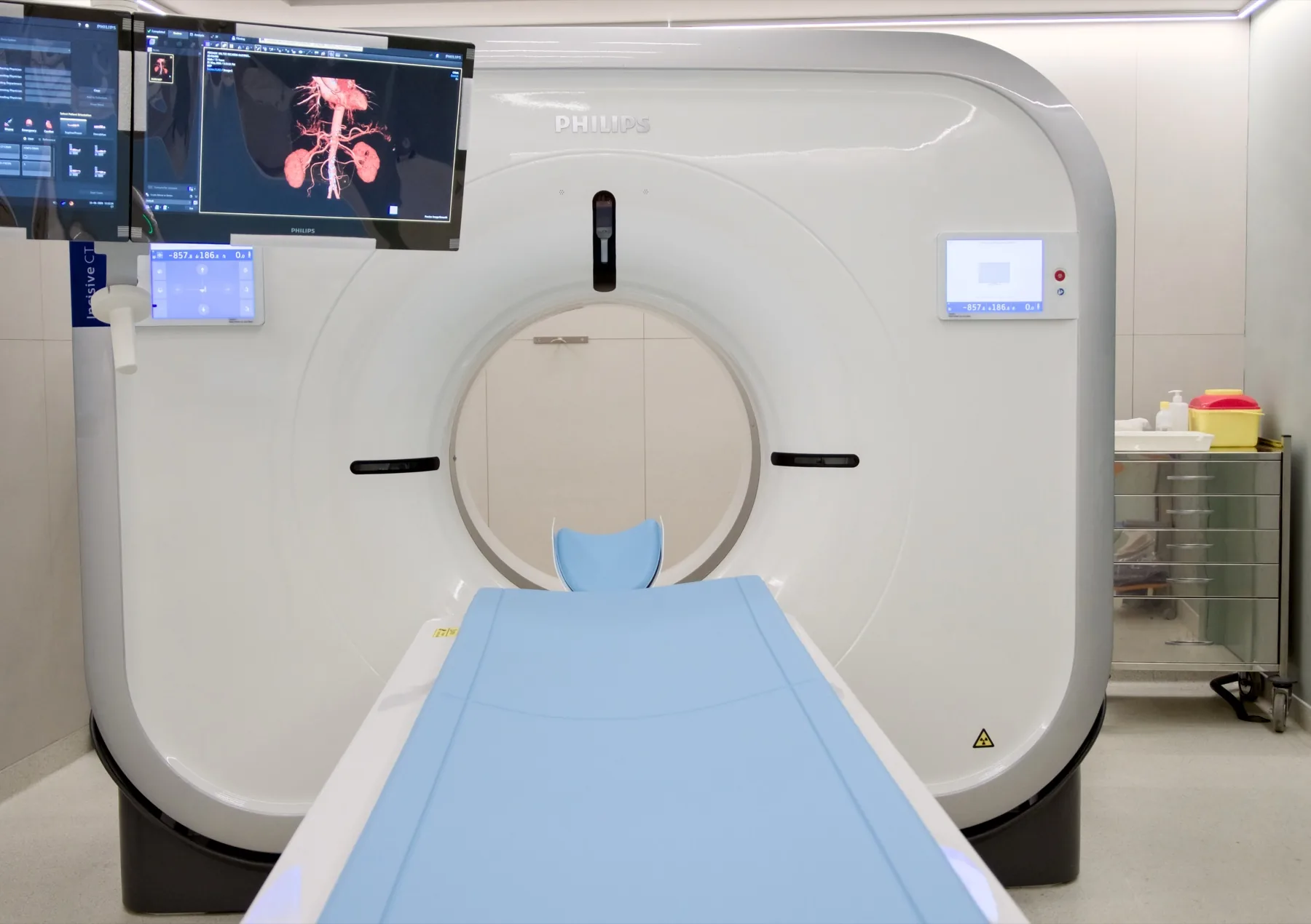
The table moves through the scanner's open ring — it is not a tunnel. You just need to keep your head still for a few brief moments.
- 5
Contrast (when indicated)
Iodinated contrast is given intravenously. It is normal to feel a passing warmth through the body and a metallic taste — these pass within a few minutes.
- 6
Finishing
You can leave, drive and resume your normal day. If you received contrast, we recommend drinking water throughout the day.
Preparation
- Fasting: not required, with or without contrast. Have a light meal before you come and drink water freely up to the time of your exam.
- Kidney function: if the exam is with contrast and you have recent blood tests (last 3 to 6 months) with creatinine/kidney function, it's advisable to bring them. They are not mandatory.
- Medication: keep your regular medication — including your diabetes and blood pressure medication, and metformin — unless your doctor advises otherwise.
- Iodinated contrast allergy: if you have had a reaction to iodinated contrast, let us know when booking. Each case is assessed individually by the radiologist.
- Metal objects: remove only those from the area being studied — hairpins, earrings, piercings, glasses, removable dentures, hearing aids.
- Clothing: as a rule, you keep your own clothes on.
- Pregnancy: CT uses X-rays. If you are or think you may be pregnant, always let us know before the exam.
- Breastfeeding: always let us know. If the exam is with contrast, breastfeeding may continue as normal after the contrast is given (ACR/ESUR recommendation).
- Children: let us know when booking, so we can prepare the exam and adjust the protocol.
- Documents: referral, insurer/scheme card (if applicable), previous head exams (CT/MRI) and recent blood tests, if you have them.
Results
The report is produced by a radiologist from the images. The report is available as a PDF (by email or printed) and the images on CD and on CRMA's imaging platform, to take to your doctor.