Mon–Fri 08:00–19:00 · Sat 08:00–13:00· Faro· +351 289 892 100· geral@crmalgarve.pt PTENFR
CRMA
Magnetic Resonance Imaging · By area

Magnetic Resonance Imaging of the Abdomen and MRCP

Allows detailed assessment of the organs of the upper abdomen — the liver, bile ducts, pancreas, spleen, kidneys and adrenal glands — with no radiation. It includes MRCP (magnetic resonance cholangiopancreatography), which studies the bile ducts without contrast or endoscopy.

Duration30–45 min
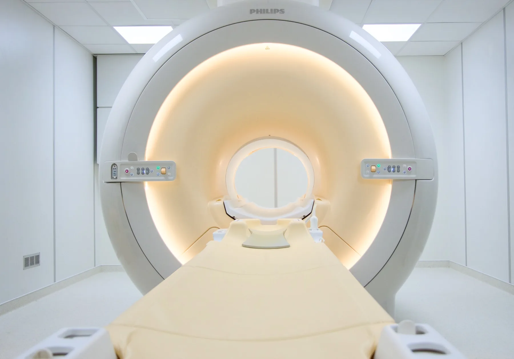
EquipmentPhilips 1.5T
ContrastSometimes
Preparation4 h fasting
CRMA's Magnetic Resonance Imaging (MRI) scanner, in Faro
In summary

An abdominal magnetic resonance (MRI) scan uses a magnetic field, with no radiation, to study the organs of the upper abdomen in detail — the liver, bile ducts, pancreas, spleen, kidneys and adrenal glands. It includes MRCP (magnetic resonance cholangiopancreatography), which assesses the bile ducts and the pancreatic duct without contrast or endoscopy. At CRMA, in Faro, it takes about 30 to 45 minutes, is done fasting and may use contrast. During the exam, you will be asked to hold your breath, on breathing out, at a few points.

What is assessed in an abdominal MRI?

Abdominal MRI allows assessment of the organs of the upper abdomen — the liver, the gallbladder and bile ducts, the pancreas, the spleen, the kidneys and the adrenal glands — and the tissues around them. It is particularly useful for characterising lesions already identified on other exams (ultrasound or CT).

MRCP (magnetic resonance cholangiopancreatography) is an MRI technique that studies the bile ducts and the pancreatic duct non-invasively — without contrast and without endoscopy — unlike ERCP (the endoscopic procedure). Because it uses a magnetic field, it involves no ionising radiation. The report is written by a radiologist; the clinical interpretation rests with your treating doctor.

In addition to the study of the upper abdomen, targeted MRI studies of a single organ may be carried out — for example, MRI of the pancreas, renal MRI or MRI of the adrenal glands — according to the clinical indication.
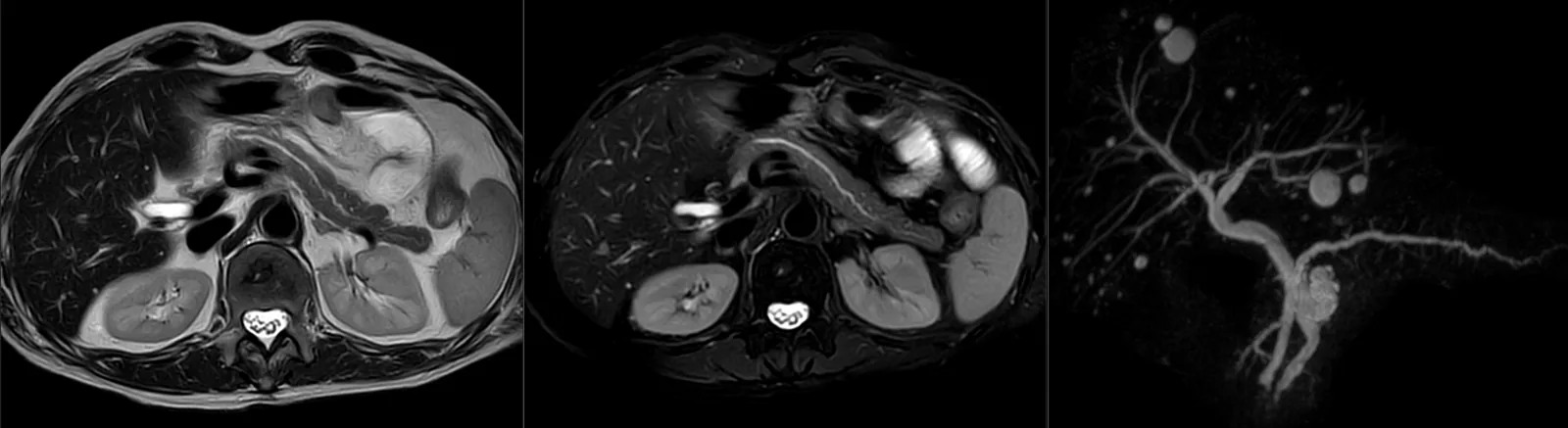
Abdominal magnetic resonance imaging and MRCP: axial slices of the upper abdomen — liver, pancreas, spleen and kidneys — and a reconstruction of the bile ducts and pancreatic duct (MRCP).

When it is requested

  • Characterisation of liver lesions (cysts, haemangiomas, nodules) seen on another exam.
  • Surveillance in chronic liver disease, according to medical guidance.
  • Study of the bile ducts (MRCP) — suspected stones in the common bile duct, dilated bile ducts or jaundice to clarify.
  • Study of the pancreas — chronic pancreatitis, lesions or cysts.
  • Characterisation of adrenal nodules.
  • Study of the kidneys and urinary tract, in specific situations.

What the exam is like

  • 1Before

    Reception and safety questionnaire

    You come having fasted for 4 hours and fill in the safety questionnaire. Note any recent kidney-function blood test, if you have one.

  • 2

    Changing room and venous access

    You remove any metal objects and put on a gown. When contrast is used, a venous line is placed in your arm.

  • 3

    Positioning

    You lie on your back, with a body coil over your abdomen.

  • 4

    Breath-holds

    At several points you will be asked to hold your breath, on breathing out, for a few seconds — this is the most important thing you can do to help produce sharp images.

  • 530–45 min

    Image acquisition

    Several sequences, including MRCP. When indicated, contrast is given, sometimes one specific to the liver.

  • 6

    Finishing up

    You get dressed and can leave without needing anyone to accompany you.

Preparation

  • Changing and clothing: as a rule, you undress and stay in underwear with no metal parts — usually pants and socks. Depending on the area, the radiographer may allow you to keep some clothing on. You are given a gown.
  • Fasting: 4 hours beforehand (it helps with the study of the bile ducts). You may drink water, unless told otherwise.
  • Contrast and kidney function: a recent kidney-function blood test may be requested; let us know of any kidney disease.
  • Breathing: the exam requires you to hold your breath (on breathing out) at a few points; let us know if you find this difficult.
  • Pregnancy: always let us know; it affects the use of contrast.
  • Documents: your referral, your insurer/scheme card (if you have an agreement), and any previous abdominal exams (ultrasound, CT, previous MRI).

Results

The report is written by a radiologist from the images. The report is available as a PDF (by email or printed) and the images on CD and on CRMA's imaging platform, to take to your treating doctor — gastroenterologist, surgeon, family doctor or other.

Frequently asked questions

About abdominal MRI and MRCP.

Does an abdominal MRI hurt?

No. It is a painless exam. When contrast is used, you only feel the pinprick of the venous line in your arm.

Do I have to fast?

Yes, 4 hours beforehand. Fasting helps above all with the study of the bile ducts (MRCP). You may generally drink water.

Will I have to hold my breath?

Yes, at several points. You will be asked to hold your breath, on breathing out, for a few seconds, so that the images of the organs are sharp. The radiographer guides you through each step.

What is MRCP?

It is an MRI technique that studies the bile ducts and the pancreatic duct without contrast and without endoscopy. Unlike ERCP (the endoscopic procedure), MRCP is not invasive.

Will I need contrast?

It depends on the indication. Some studies are done without contrast (for example, MRCP on its own); others use intravenous contrast, sometimes one specific to the liver.

How long does it take?

In general, between 30 and 45 minutes, depending on what is studied and whether contrast is used.

Can I have an abdominal MRI if I'm pregnant?

Always let us know of a confirmed or possible pregnancy. MRI does not use ionising radiation and may be performed at any stage of pregnancy when the doctor considers the information necessary; in the first trimester, as a precaution, the indication is weighed case by case. Contrast (gadolinium) is avoided throughout pregnancy unless clinically necessary.

What's the difference between abdominal MRI and CT or ultrasound?

Ultrasound is fast and often the first exam; CT uses X-rays and is very useful in a range of situations; MRI gives great detail of the soft tissues and the bile ducts, without radiation. They are complementary exams.

Ready to book?

Book your abdominal MRI or MRCP at CRMA.

In Faro, with personal service in PT, EN and FR. We confirm your booking and give you the fasting and preparation instructions.