
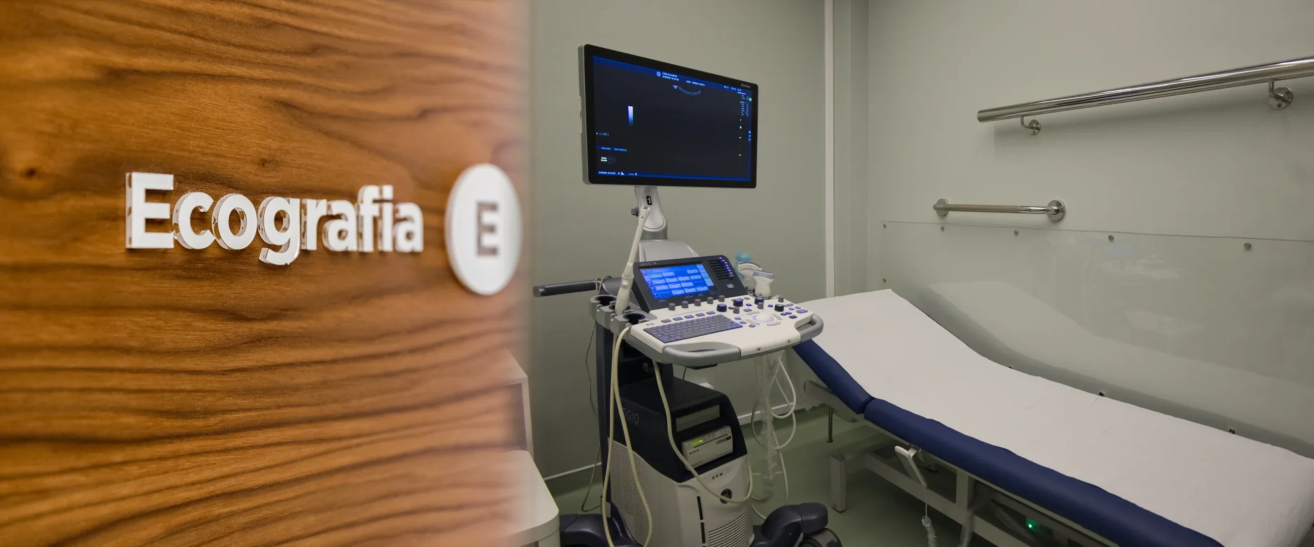
What can be assessed with an ultrasound?
Ultrasound allows assessment of a wide range of organs and areas. At CRMA we perform, among others, the following groups of studies:
- Abdomen and urinary tract — liver, gallbladder, pancreas, spleen, kidneys, bladder; includes assessment of the post-void residual.
- Prostate — by suprapubic or transrectal approach, depending on the referral.
- Pelvic and gynaecological — by suprapubic or endocavitary approach.
- Obstetric — pregnancy monitoring, including the second and third trimesters and twin pregnancy.
- Thyroid, parathyroid glands, salivary glands and soft tissues of the neck.
- Breast — on its own or as a complement to mammography.
- Musculoskeletal and soft tissue — muscles, tendons and joints.
- Scrotal.
Because it uses ultrasound rather than X-rays, this exam involves no ionising radiation, so it can be repeated whenever clinically necessary and used safely during pregnancy. The Doppler study (assessment of the vessels and blood flow) is a complementary technique with its own page.
When it is requested
- Abdominal or pelvic pain in initial assessment.
- Assessment of urinary complaints — kidneys, bladder, prostate.
- Pregnancy monitoring.
- Palpable lumps — thyroid, breast, soft tissue.
- Muscle, tendon or joint pain.
- Follow-up of already known findings, as directed by your treating doctor.
The exam is always requested by the referring doctor. For a check-up without a referral, see the Prevention page — the indication is taken on by a CRMA radiologist, after a prior assessment.
What the exam is like
- 1Before
Reception
Arrive at the time given when booking, with your referral and any previous exams, if you have them.
- 2
Preparation in the room
Depending on the area, you may be asked to uncover only the region to be studied. For endocavitary studies, the team explains the procedure and ensures your privacy.
- 3
Gel and probe
The doctor applies a water-based gel to the skin and slides the probe over the area, viewing the images in real time. The gel wipes off easily afterwards.
- 45–25 min
Image acquisition
You may be asked to make small changes of position or to hold your breath to improve the view.
- 5
Finishing up
The gel is wiped off and you can carry on with your day with no restrictions.
Preparation
Preparation depends on the type of ultrasound and is always confirmed when you book. As a rule:
- Abdominal ultrasound: fast for 4 to 6 hours, depending on the study and on the instructions given when booking. You may take your regular medication with a little water up to 1 hour before the exam. If you have diabetes: do not take your diabetes medication on an empty stomach (you may take the rest up to 1 hour before, with a little water).
- Bladder, pelvic or prostate ultrasound by the suprapubic approach: this is done with a full bladder. How much water to drink and when depend on the type of exam and are given when booking.
- Renal ultrasound: no preparation.
- Endovaginal gynaecological ultrasound: no preparation; it should not be performed during your period.
- Transrectal prostate ultrasound: may require an enema (in the morning, on waking) — confirmed when you book.
- Obstetric, thyroid, breast, musculoskeletal, soft-tissue and scrotal ultrasound: no preparation.
- Children: preparation may differ from the usual and is given when you book.
Results
The report is written by a radiologist. The report is available as a PDF (by email or printed) and the images on CRMA's imaging platform, to take to your treating doctor. With written authorisation, they can be sent directly to the referring doctor.