What MR angiography is
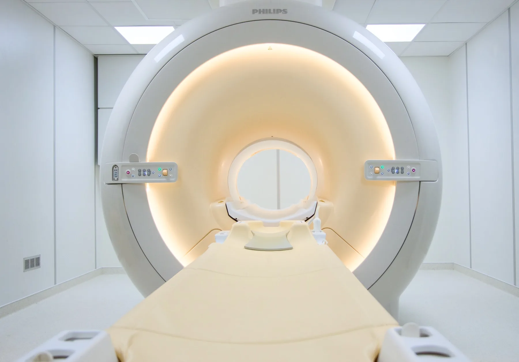
MR angiography (magnetic resonance angiography) allows the arteries and veins to be visualised using the magnetic field of the MRI scanner, without X-rays. It differs from CT angiography in three respects: it uses no ionising radiation; it can be performed with or without contrast (Group II macrocyclic gadolinium); and in some territories it is more sensitive, particularly for arteriovenous and soft-tissue malformations.
When it is requested
- Known or suspected vascular disease: atherosclerotic stenoses (carotid, renal, peripheral), aneurysms, dissections, thrombosis.
- Vascular malformations: cerebral or peripheral AVMs, dural arteriovenous fistulas (cerebral and spinal), soft-tissue vascular malformations.
- Venous thrombosis: cerebral venous thrombosis (dural sinuses), atypical DVT, May-Thurner syndrome, pelvic varices.
Arterial MR angiography
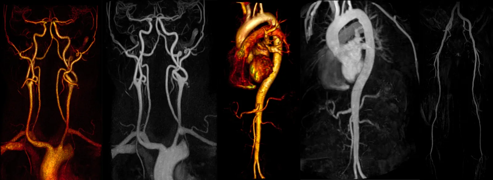
We perform it using non-contrast Time-of-Flight (ToF) (the first choice for the circle of Willis and neck vessels) or CE-MRA with Group II macrocyclic gadolinium (the standard for the thoracic and abdominal aorta, renal, iliac and peripheral arteries).
Territories available: head (circle of Willis), neck vessels (carotid and vertebral), pulmonary, thoracic and abdominal aorta, renal and iliac arteries, lower limbs (runoff) and upper limbs, face.
Venous MR angiography
Cerebral venography is the first-choice exam for suspected thrombosis of the dural venous sinuses — an uncommon but important cause of persistent headache. We perform 2D-TOF, non-contrast 3D-PC, and CE-MRV with gadolinium in selected cases.
Other venous territories available: neck, abdominal, pelvic (varices), lower limbs (May-Thurner) and upper limbs (venous thoracic outlet syndrome).
4D time-resolved MR angiography
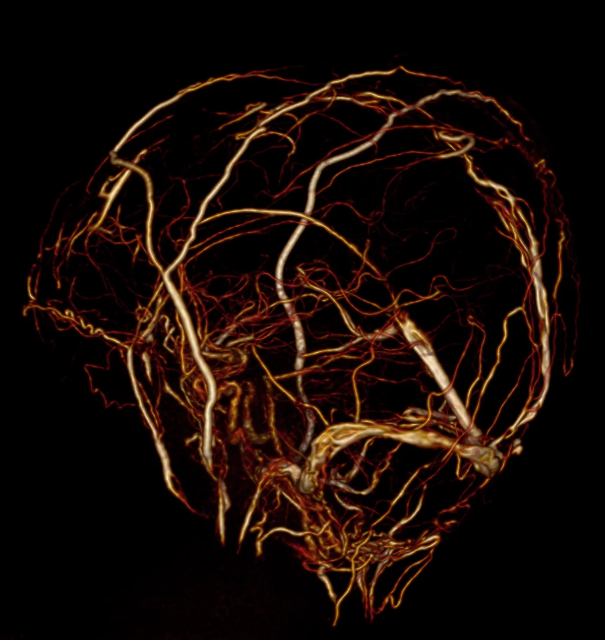
An advanced technique with high temporal resolution — it acquires several images per second, allowing the passage of contrast through the vessels to be observed in successive phases. It is particularly useful in cerebral arteriovenous malformations (AVMs) (it identifies early draining veins, a pathognomonic sign), cerebral and spinal dural arteriovenous fistulas, soft-tissue vascular malformations and pelvic varices.
CRMA performs 4D MR angiography of the head, face and extremities. In many cases it can avoid the need for invasive digital subtraction angiography (DSA).
What the exam is like
- 1Before
Reception and safety questionnaire
Arrive 15 minutes early and fill in the safety questionnaire, if you haven't already done so online.
- 2
Changing room
You undress down to underwear with no metal parts; you are given a gown.
- 3
Cannula for contrast (when applicable)
If the exam includes contrast (CE-MRA, 4D), a cannula is placed in your arm and removed at the end.
- 4
Positioning
You lie on your back; the coil used depends on the territory — head, neck, chest or limbs.
- 5
Inside the scanner
Depending on the territory, your head or trunk is inside the magnet. You stay in constant contact with the radiographer.
- 625–50 min
Image acquisition
Sequences with a characteristic knocking noise; you are given ear protection. When contrast is used, the injection is timed with the acquisition — you may feel a cool sensation in your arm. For some sequences you are asked to hold your breath briefly.
- 7
Finishing up
The cannula is removed. After contrast, drinking extra fluids in the following hours is recommended.
Preparation
- Fasting: not needed for studies of the head, neck and limbs, even with contrast. 4 hours' fasting still applies to abdominal MR angiography (abdominal aorta and renal arteries) — the instruction is confirmed when you book.
- Kidney function: if you have known kidney disease, tell us when you book. The contrast agents used (gadolinium-based, Group II macrocyclic) have a strong safety profile; assessment is case by case.
- Allergies: any previous reaction to gadolinium should be reported when you book.
- Pregnancy and breastfeeding: always tell us. Breastfeeding may continue as normal after a Group II macrocyclic gadolinium agent (ACR/EMA recommendation).
- Claustrophobia: for cerebral and cervical MR angiography your head is inside the magnet. If you are anxious about this, contact us before the day of the exam.
Results
The report is written by a radiologist, based on the images and the vascular reconstructions (MIP, MPR, VR). The report is available as a PDF (by email or printed) and the images on CD and on CRMA's imaging platform. Where urgent findings are suspected, CRMA's radiologist contacts the referring doctor directly.