
What is Magnetic Resonance Imaging?
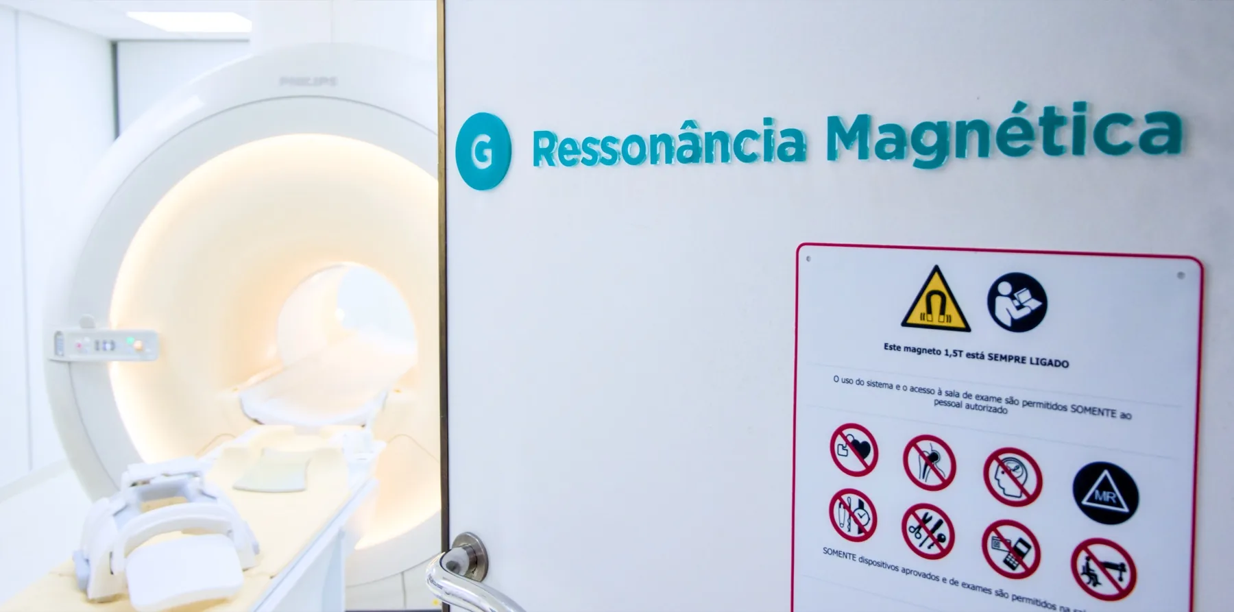
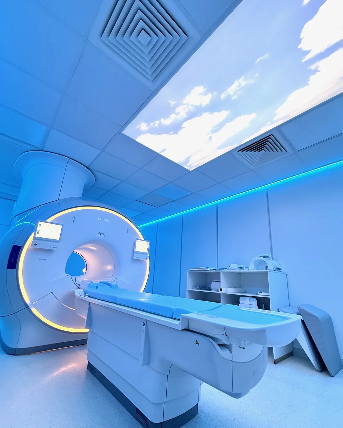
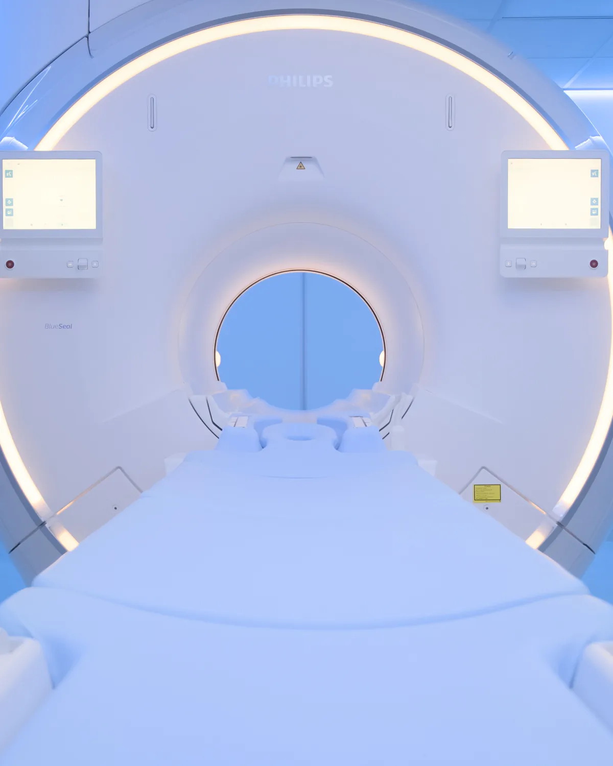
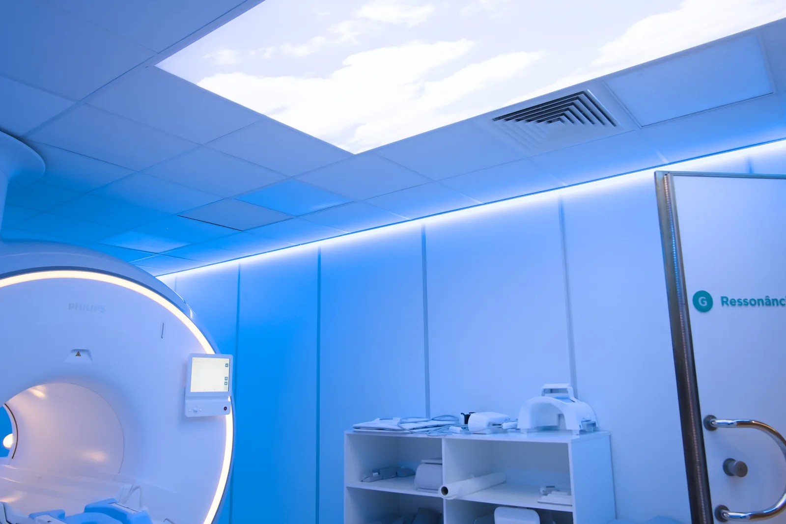
Magnetic resonance imaging (MRI) is a medical imaging technique that uses a strong magnetic field and radio waves to produce detailed images of the inside of the body. Unlike CT and X-rays, it uses no ionising radiation.
It is often requested to study soft tissues that other exams struggle to show in the same detail: the brain, spinal cord, joints, blood vessels, abdominal and pelvic organs, breast and reproductive system.
The images are read and reported by a radiologist. In some cases, intravenous contrast (gadolinium) is given to improve visualisation. The contrast agents used at CRMA are ACR Group II macrocyclic agents, with a high safety profile even in patients with impaired kidney function.
How it's done
The exam usually takes between 20 and 60 minutes, depending on the region to be studied and the type of MRI. It is carried out by a radiographer, following a protocol set by the radiologist.
- We recommend arriving 15 minutes before your appointment time.
- You should fill in the safety questionnaire beforehand.
- You remove all metal objects in the changing room.
- The exam is performed lying on your back, in constant contact with the radiographer.
- The equipment makes a characteristic noise. In some exams we can reduce it, and you are always given ear protection.
- For some sequences, intravenous contrast is given through a cannula in the arm.
Preparation
Preparation varies depending on the region to be studied and whether or not contrast is used. Specific instructions are always given when you book. In general:
- Fasting: as a rule, not required — contrast (gadolinium) does not call for fasting. Four hours of fasting still applies only to some upper abdomen studies (liver and bile ducts, MRCP, pancreas, kidneys and adrenal glands, abdominal MR angiography) and 4 to 6 hours to MR enterography, for which it is also best not to drink water in the 2 to 4 hours beforehand. The instruction is confirmed when you book.
- Clothing: comfortable, with no metal parts. You may be given a gown.
- Metal objects: remove them all — jewellery, piercings, watches, glasses, removable dentures, cards with a magnetic strip.
- Documents: your referral, your insurer or subsystem card, and any previous exams.
Results
The report is written by a radiologist based on the images. The report is available as a PDF (by email or printed) and the images on CD in DICOM format and on CRMA's imaging platform, to take to your doctor. We let you know when it is ready, as agreed when you booked.
Variants of Magnetic Resonance Imaging
Eleven MRI exams have their own page, with preparation, step-by-step details and frequently asked questions:
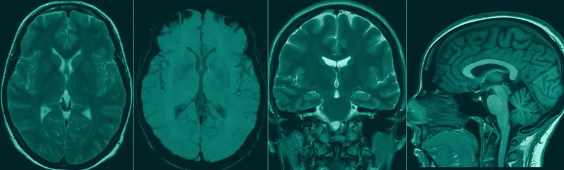
Head MRI
The brain and intracranial structures.
Learn more →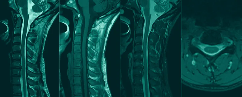
Cervical Spine MRI
Vertebrae, discs and the cervical spinal cord.
Learn more →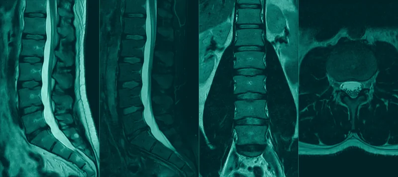
Lumbar Spine MRI
Discs, vertebrae and the lumbar nerve roots.
Learn more →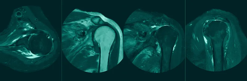
Shoulder MRI
Rotator cuff, instability and surrounding tissues.
Learn more →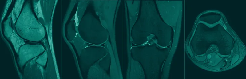
Knee MRI
Menisci, ligaments and cartilage.
Learn more →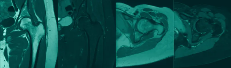
Hip MRI
The hip joint and surrounding tissues.
Learn more →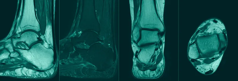
Foot and Ankle MRI
Tendons, ligaments and the plantar fascia.
Learn more →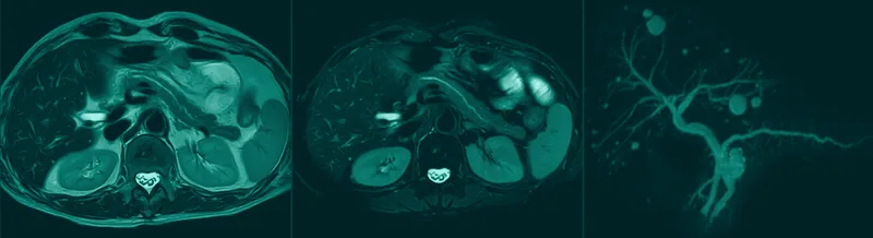
Abdominal MRI
Liver, bile ducts (MRCP), pancreas and kidneys.
Learn more →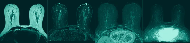
Breast MRI
Complementary study of the breast.
Learn more →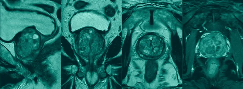
Prostate MRI
Multiparametric study of the prostate.
Learn more →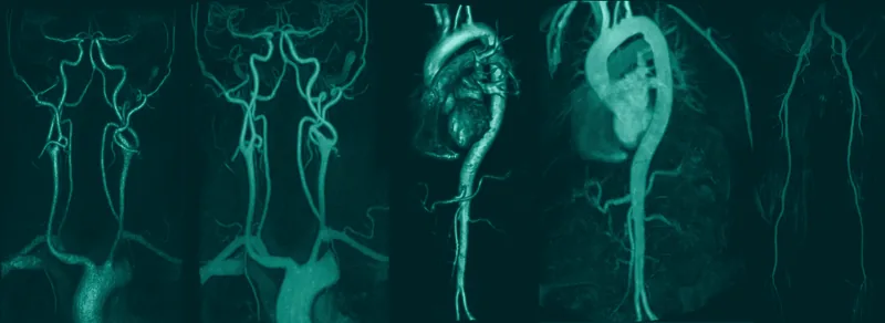
MR angiography
Vascular studies: arterial, venous and 4D time-resolved.
Learn more →Full list of variants by region — each exam is performed with a dedicated protocol, adjusted by the radiologist to the clinical indication:
- Head and nervous system: head, pituitary gland, orbits, ear, paranasal sinuses, face, temporomandibular joints.
- Spine: cervical, thoracic, lumbosacral, coccygeal, spinal cord.
- Joints and limbs: shoulder, elbow, wrist, hand, hip, knee, foot, ankle.
- Chest, abdomen and pelvis: chest, breast, abdominal (with MRCP and MR enterography), pancreas, adrenal glands, renal, pelvic, uterus and ovaries, anal and perianal fistulas, multiparametric prostate.
- Vascular studies: arterial, venous and 4D time-resolved MR angiography (for vascular malformations).