
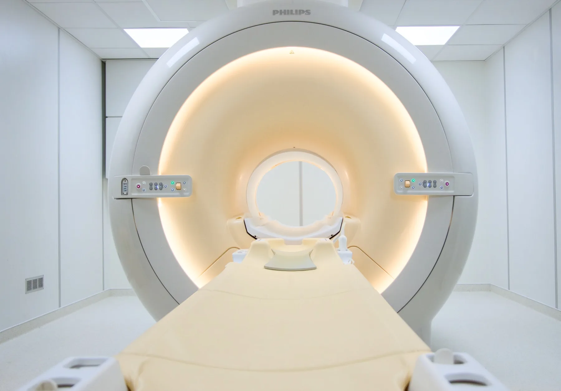
What is assessed in a breast MRI?
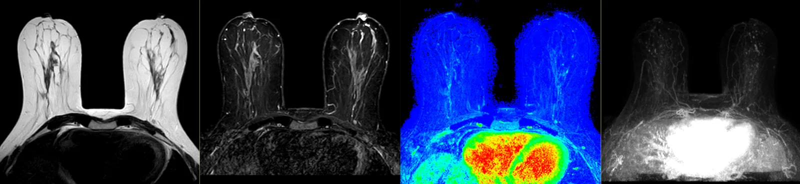
Breast MRI allows assessment of the tissue of both breasts in high detail, including the chest wall, and helps characterise areas identified on other exams (mammography or ultrasound). It also allows assessment of the integrity of breast implants.
Because it uses a magnetic field, it involves no ionising radiation. It is a highly sensitive exam, usually performed with intravenous contrast (gadolinium). It is important to understand that breast MRI complements — it does not replace — mammography and ultrasound. The choice and reading of exams are the doctor's responsibility; the report is written by a radiologist.
When it is requested
The referral always comes from your doctor. These are the situations in which it is usually requested, always as a complement to other exams:
- Monitoring of women at high risk of breast cancer (e.g. carriers of a mutation such as BRCA, or a significant family history).
- Characterisation of findings on mammography or ultrasound that need further information.
- Assessment of the extent of a known disease, before a treatment decision.
- Assessment of the response to treatment (e.g. chemotherapy before surgery).
- Assessment of breast implants (e.g. suspected rupture).
- Clarification of discrepancies between exams.
What the exam is like
- 1Before
Reception and safety questionnaire
Arrive 15 minutes early and fill in the safety questionnaire. Let us know if you have a recent kidney-function blood test, because of the contrast.
- 2
Changing room and venous access
You undress from the waist up and put on a gown. When contrast is used, a venous line is placed in your arm.
- 3
Positioning
You lie face down on a table with dedicated openings, where the breasts hang within a dedicated coil, with your arms above your head.
- 4
Inside the scanner
The table moves into the scanner. You stay in contact with the radiographer and are given an emergency call button.
- 520–40 min
Acquisition with contrast
Sequences are acquired before and after the contrast, to study how the tissue takes it up over time. It is important to stay still.
- 6
Finishing up
The venous line is removed; you get dressed and can leave without needing anyone to accompany you.
Preparation
- Fasting: not needed, even with contrast. Have a light meal before you come and drink water freely.
- Contrast and kidney function: usually with gadolinium (macrocyclic); a recent kidney-function blood test may be requested.
- Pregnancy and breastfeeding: always let us know — it affects the use of contrast.
- Changing room and clothing: you undress from the waist up and put on a gown; you keep your lower underwear. No metal parts (including underwired bras); remove jewellery.
- Documents: your referral and previous breast exams (mammography, ultrasound, previous MRI), important for comparison.
Results
The report is written by a radiologist, often compared with your previous exams. The report is available as a PDF (by email or printed) and the images on CD and on CRMA's imaging platform, to take to your treating doctor (breast specialist, gynaecologist, surgeon, oncologist or family doctor).